
One of the efforts to support the productivity of Bali cattle is to utilize ovaries from the slaughterhouse by using assisted technology, namely in vitro embryo production. This study aims to determine the developmental ability and to measure the concentration of hydrogen peroxide (H2O2) in Bali cattle embryos produced in vitro. 542 oocytes were matured in TCM 199 medium, were fertilized and were cultured using CR1aa medium for 48 h. After 48 h of culture, the embryo developed into 2, 4, 8 and 16 cells. Furthermore, it was grouped based on the stage of development, each of which was re-cultured for 48 h. After that, its development was re-evaluated and H2O2 measurements were taken. The 2-cell embryo was not able to reach the morula, while stage 4-, 8- and 16-cell were able to develop to the morula stage as much as 6(9.83%), 5(12.83%) and 4(25%), respectively. The concentration of H2O2 in each group of cells did not differ significantly (P>0.05). However, the results of the correlation test between the H2O2 concentration and the embryo’s ability to reach the morula stage were negatively correlated. The lower the concentration of H2O2, the higher the chances of the embryo reaching the morula stage. The conclusion is that the high H2O2 can inhibit the development of Bali cattle embryos that are cultured in vitro.
Research Article · Volume 55, Issue 6, 30 October 2023 · Open Access
Developmental Ability and Concentration of Hydrogen Peroxide in Bali Cattle Embryos Cultured In Vitro
· · ·
Affiliations & Notes ⌄
- 1Agricultural Science Study Program, Graduate School, Hasanuddin University, 90245 Makassar, Indonesia 2Department of Animal Production, Faculty of Animal Science, Hasanuddin University, 90245 Makassar, Indonesia * Corresponding author: sonjayaherry@gmail.com
Article Info ⌄
- Journal:
- Pakistan Journal of Zoology
- Published:
- 30 October 2023

From the article
- The developmental ability of Bali cattle embryos 48 h after culture varies, namely 2-,4-,8- and 16-cell.
- Embryos cultured back from 4-cell 8-cell and 16-cell for 48 h were able to reach the morula.
- The results showed that there was no significant influence (P>0.05) between each group of embryonic cells which was re-cultured based on its cell count group and the H2O2 concentration levels in Bali cattle embryos.
- The H2O2 relationship forms a negative trend, indicating that the lower the concentration of H2O2 in the embryo, the higher the embryo’s ability to reach the morula (Fig. 4).
Selected from the article's own text.
Abstract
INTRODUCTION
Bali cattle are native Indonesian cattle widely developed by the farming community in South Sulawesi. One way to continue supporting the productivity of Bali cattle is to utilize ovaries from slaughterhouses by using assisted reproductive technology, namely in vitro embryo production. In general, studies on embryo production have been widely conducted, but those carried out on Bali cattle are still limited in number. In vitro embryo production is still being improved to support the success of embryos that are worthy of transfer . The initial development embryo often experiences obstacles and becomes a critical problem for embryo production, resulting in obstacles in the production of embryos in vitro . Several factors can affect the successful production of embryos in vitro including donors, sperm, heredity and the environment . In addition, different culturalsystems can also affect embryonic development . In vitro production of Bali cattle embryos resulted in zygote cultures which show varying rates of embryonic cell division indicated by the occurrence of 2-, 4-, 8- and 16-cell embryos on the second day or 48 h after culture ; . The in vitro embryo culture system has not been able to be properly conditioned to resemble in vivo so that it can lead to the increase of reactive oxygen species .
Reactive oxygen species (ROS) are oxygen-derived molecules that act as strong oxidants. ROS produces oxygen free radicals such as hydrogen peroxide (H2O2), hydroxyl radicals (OH) and superoxide anion radicals (O2), and is a by-product of cell metabolism . Small amounts of ROS are needed in some physiological processes such as maintaining embryonic development , and a small amount of H2O2 can maintain the balance of cell mass , spermatozoa capacity and acrosome reactions . An increase in ROS at the intracellular level can cause developmental disorders, apoptosis and embryo death ; . Oocytes that experience oxidative stress can cause problems during meiosis and increase fragmentation in the embryo . ROS is also an obstacle to the glycolysis process so the embryo does not optimally utilize glucose as an energy source .
The success of in vitro embryo production can be judged by the number of embryos that are able to develop after the fertilization process . The success of embryo culture in vitro is thought to be related to H2O2 concentrations, high H2O2 concentrations can inhibit embryo development. Information regarding the measurement of H2O2 in Bali cattle embryos produced in vitro does not yet exist. This study focuses on measuring the concentration of H2O2 in Bali cattle embryos based on cultures with the same number of cells.
MATERIALS AND METHODS
Oocyte collection and selection
This study used 138 pairs of ovaries with 542 oocytes obtained from slaughterhouse. The cattle ovaries were obtained from slaughterhouse and brought to the laboratory by transport media (0.9% NaCl solution). The oocytes collection was carried out using the slicing method and then were selected using the Olympus SZ51 Japan microscope (only Grade A and B oocytes were used). Grade A has a compact cumulus-oocyte complex (COC) cell surrounded by at least 4-5 layers of cumulus cells and homogeneous cytoplasm. Meanwhile, grade B has a COC surrounded by 2-3 layers of cumulus cells attached to the zona pellucida and homogeneous cytoplasm .
Oocyte maturation
The selected oocytes were then washed with phosphate buffered saline (PBS) (Gibco by life technologies, USA) added with 0.2% bovine serum albumin (BSA) (Sigma-Aldrich, USA) matured in a maturation medium consisting of medium 199 (Gibco by life technologies, USA) added with 0.3% BSA, 10 IU/ml Pregnant Mare Serum Gonadotropin (PMSG) (Intergonan, Intervet Deutschland GmbH), 10 IU/ml human chorionic gonadotropin (hCG) (Chorulon, Intervet international BV Boxmeer-Holland), and 50 μg/ml gentamycin (Sigma-Aldrich, USA). Maturation was conducted in the form of drops (80 μl / drop) containing 10-15 per drop and covered with mineral oil (Sigma Chemical Co. St. Louis MO, USA). Maturation was carried out in a 5% CO2 incubator with a temperature of 38.5 oC for 24 h . In this study, oocytes that experienced an expansion of cumulus cells or activation of cumulus cells, were used as an indication of oocyte maturity and continued for fertilization.
In vitro fertilization
The semen used was obtained from the same bull. It was thawed for 20 seconds at 37 oC, was centrifuged for 5 min at 1500 rpm 2 times, split the supernatant and spermatozoa, and then semen was added with fertilization media so that the final concentration of spermatozoa was 1.5×10⁶ cells/ ml ; . After that, prepared four drops on a petri dish (80 μL/drop) covered by mineral oil (Sigma Chemical Co. St. Louis MO, USA), equilibrated for 30 min. The matured oocytes were washed 2 times, put into IVF media and stored in a 38.5oC incubator and a 5% CO2 concentration for 5-6 h.
In vitro culture
Oocytes after 5-6 h of fertilization, the oocytes were washed twice using CR1aa culture medium, then transferred in a drop of 80 μl CR1aa culture medium (modification by ; added with 5 mg/ml of BSA, 2.5% FBS and covered by mineral oil (Sigma Chemical Co. St. Louis MO, USA), which then was cultured in an incubator of 5% CO2 at 38.5°C (Meo et al., 2005). The evaluation of embryo division ability is carried out 48 h after culture, then the embryos that develop are re-cultured and as a control, namely embryos that develop are cultured without grouping the number of cells and grouped based on the stages of division, namely embryo 2, 4, 8, and 16-cells. Then the embryo is re-cultured for 48 h and re-evaluated.
Hydrogen peroxide (H2O2) measurements
Embryos that have been re-cultured for 48 h, namely control and embryos that have been grouped based on the stages of division, then concentration measurements are carried out H2O2 concentration was measured using 2’,7’-dichlorodihydro fluorescein diacetate (DCHFDA) (Sigma) as described by Gustina et al. (2019) with minor modifications. Embryos in each group were incubated for 15 min with a culture medium containing 10 μM of DCHFDA, were washed in a fresh medium before placed on a glass slide and were covered by a cover glass. Fluorescence emission was recorded with a digital camera (Zeiss AxioCam HRc, Germany) which was attached to a fluorescence microscope (Zeiss Axio Imager A2) after excitation at 480 nm and emissions of 510 nm. Fluorescent images were converted into TIFF files using Adobe Photoshop CS3 (Adobe Systems Inc., San Jose, CA), then were analyzed using ImageJ 1.47 software (Sun Microsystems, Inc., California, USA). Fluorescent imagery was measured by counting the number of pixels after color reversal. Fluorescence intensity represents intracellular H2O2 concentration.
Data analysis
The data of embryo developmental ability were analysed descriptively, the H2O2 concentrations were analysed by one-way analysis of variance (ANOVA) and the correlation test was conducted using SPSS 20.00 and significance level of 0.05.
RESULTS
The developmental ability of Bali cattle embryos 48 h after culture varies, namely 2-,4-,8- and 16-cell. Once grouped by the cleavage stages and re-cultured for 48 h, the embryos of Bali cattle that can develop further also vary, as presented in Figure 1. The total number of embryos cultured was 228 embryos consisting of control or no grouping (48 embryos), 2-cells (64 embryos), 4-cells (61 embryos), 8-cells (39 embryos) and 16-cells (16 embryos). The group of embryos cultured of 2-cell was able to develop up to 16-cell by 3.27%. Embryos cultured back from 4-cell 8-cell and 16-cell for 48 h were able to reach the morula. The number of embryos that reached the morula was 15 embryos. Overall, the data of embryo development cultured from a different number of cells for 48 h and continued to be re-cultured for 48 h is presented in Figure 1.
Evaluation of H2O2 concentration was carried out by measuring the intensity of H2O2 concentration in Bali cattle embryos (Fig. 2). The brighter the green color, the higher the concentration of H2O2 in Bali cattle embryos. The results showed that there was no significant influence (P>0.05) between each group of embryonic cells which was re-cultured based on its cell count group and the H2O2 concentration levels in Bali cattle embryos. The concentration of H2O2 in the control group was 168.08±58.77, while the concentration in the 2-cell embryo was 167.88±50.37, 4-cell embryo was 167.20±44.88, 8-cell embryo was 160.68± 45.78 and 16-cell embryo was 150.05±48.75 (Fig. 3).
The testing result on the relationship between the H2O2 concentration and the ability of the embryo to reach the morula stage are determined based on the results of the correlation analysis (Fig. 4). The H2O2 concentration and embryo developmental ability to reach the morula stage were negatively correlated (-0.916) and had a very strong relationship. The H2O2 relationship forms a negative trend, indicating that the lower the concentration of H2O2 in the embryo, the higher the embryo’s ability to reach the morula (Fig. 4).
DISCUSSION
In the initial development of Bali cattle embryos cultured for 48 h, there were 2-, 4-, 8 and 16-cell cleavage and 42% of which developed from 542 oocytes. The development of Bali cattle embryos cultured in vitro is different from that reported by Martins et al. (2018) who stated that the stage of cattle embryo cell development on the second day only reaches 4-cell division. Marsico et al. (2019) stated that the initial stage of embryonic development is difficult to be carried out because they involve processes that are molecularly dynamically arranged and undergo complex structural changes. The results of grouping after reculturing for 48 h showed that the 2-cell group was unable to develop until morula (Fig. 1A). This indicates that post-fertilization embryo culture is a critical period that determines the rate of division (Mizobe et al., 2017; and embryonic quality . It is further explained by Milewski et al. (2018) that most failures of in vitro embryo development occur between the 2-cell and the blastocyst stage. One of the causes of Bali cattle embryos not being able to develop is the high concentration of H2O2. It is part of ROS that can lead to lipid peroxidation and enzyme inactivation, resulting in cell damage by encouraging the formation of hydroxyl radicals . H2O2 contained in Bali cattle embryo cells is the cause of the lack of division in cells. Undeveloped embryo cells occur due to ROS production that exceeds cellular defences resulting in oxidative damage to biomolecular substances such as lipids, proteins, carbohydrates and nucleic acids, consequently inducing structural and functional changes such as lipid hydroperoxide, protein carbonylation and DNA with oxidized bases (7, 8 dihydro-8-oxoguanine) . Mitochondrial DNA (mtDNA) is also known to become more sensitive to oxidative stress . Oxidative damage to mtDNA can cause mutations and alter mitochondrial function and integrity . The results of the study of Tatemoto et al (2000) on pig oocytes cultured without cumulus cells are particularly susceptible to oxidative stress and the incidence of degeneration, the duration of DNA migration, and caspase-3 activity increased significantly in DO (denuned oocyte) exposed to ROS compared to DO cultured without XOD (xanthine oxidase).
The 4-, 8- and 16-cell group showed their ability to reach the morula (Fig. 1B, C, D). Poor embryonic development at the beginning of the culture identifies that the embryo is difficult in reaching the next stage of development. Lee et al. (2012) stated that the speed of early embryo division can be used as an indicator to assess the quality of embryos. It is further believed that cattle embryos undergo genome activation starting at stages 8-16 cell and 8-cell , so embryo development up to stage 8-cell depends on the maternal genome carried by oocytes. The process of degeneration in the early development of the embryo is likely due to drastic changes in the synthesis of embryonic proteins, internal signals that turn off maternal control in the process of embryo division and growth control, which may cause the embryo unable to be saved and eventually experience death . The development of embryo in vitro 2-cell stage that reaches the blastocyst is influenced by the outside environment and suboptimum cultural conditions to support its development . Culture media has a great contribution in the early embryonic development, the quality of blastocyst and the number of embryo cells .
The concentration of H2O2 formed in the embryo depends on high oxygen exposure to the cell. Proper concentration of H2O2 is needed to keep sperm in cattle normally functioning . However, high levels of H2O2 can lead to sperm dysfunction . Specifically, ROS serves as an intracellular signalling molecule in embryo metabolism . ROS induces lipid peroxidation in membranes, DNA damage to oocytes and is thought to cause damage to cell division, metabolite transport as well as mitochondrial function .
H2O2 concentrations have a very strong correlation (Fig. 4) with the embryo’s ability to develop to the morula stage. The higher the H2O2 content in Bali cattle embryos, the lesser the embryo’s ability to develop further. Oxidative stress, a cellular condition caused by the accumulation of H2O2, is thought to contribute significantly to the development of damaged embryos ; . Excessive levels of ROS or H2O2 are one of the main determinants of the quality of embryos produced in vivo and in vitro, caused by exposure to high oxygen concentrations, light and increased concentrations of metabolites and substrates . Oxidative stress caused by the high level of ROS production results in a reduction in ATP production and not only damages the embryo’s development but also triggers apoptotic degeneration . Excessive oxidative stress or insufficient antioxidant protection in human oocytes and embryos can cause adverse effects on male and female reproduction . Oocytes with H2O2 of more than 90 ng/oocytes undergo apoptosis (Tripati et al., 2009). Additional 30 μM H2O2 to mouse culture medium to induce oxidative stress may reduce mitochondrial membrane potential and decrease mitochondrial activity by up to 40% . An increase in H2O2 can be detrimental not only to the success rate of inductive assisted reproduction but also result in epigenetic and genetic changes in the embryo, which have an effect on transgeneration . Stress tolerance after H2O2 exposure is not mediated by the use of antioxidants such as glutathione melantonin , sericin able to block ROS . The weakness of this study is that the concentration of H2O2 in oocytes after ripening and the concentration of H2O2 in Bali cattle sperm have not been measured. It is suggested that it can be a follow-up research for Bali cattle production in vitro.
CONCLUSION
The high H2O2 can inhibit the development of Bali cattle embryos that are cultured in vitro.
ACKNOWLEDGMENTS
This research was supported and funded by the Ministry of Research, Technology and Higher Education of Indonesia through PMDSU scholarships (Stipulation Agreement No. 1343/D3/PG/2018) and research grants (Decree No. 57/UN4.22/PT.01.03/2020 with the agreement/contract number 7/AMD/EI/KP.PTNBH/2020).
Statement of conflict of interest
The authors have declared no conflict of interest.
Figures
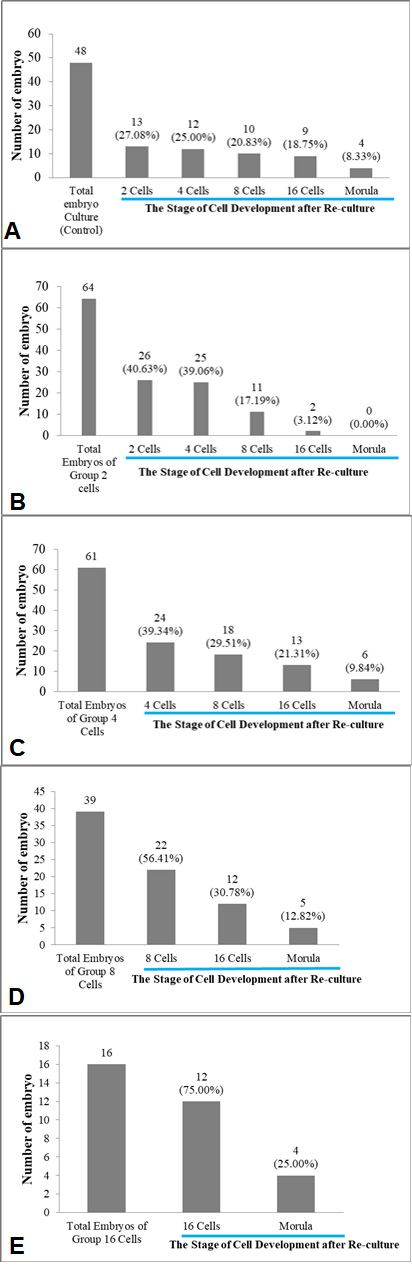
The cleavage of Bali cattle embryo after re-culture for 48 h; A, Grup of control; B, 2-cell stage (%); C, 4-cell stage (%); D, 8-cell stage (%); E, 16-cell stage (%).
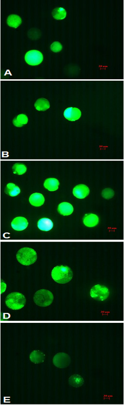
Fluorescent photomicrographs, 200× magnification. A, group of control; B, 2-cell; C, 4-cell; D, 8-cell; E, 16-cell.
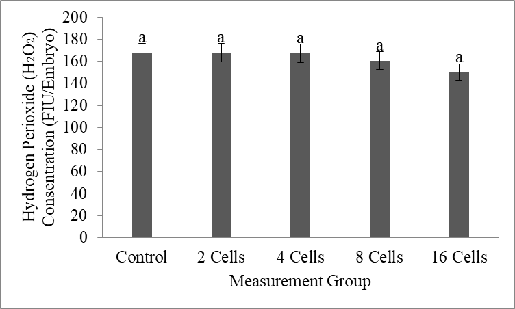
H2O2 concentration of Bali cattle embryos after re-culture for 48 h. FIU values represent fluorescence intensity units.
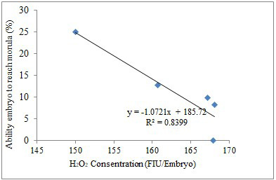
The correlation between H2O2 concentration and embryo developmental ability to reach morula stage after re-culture for 48 h (r=0.916).
References
- Abd El-Aziz, A.H., Mahrous, U.E., Kamel, S.Z., and Sabek, A.A., 2016. Factors influencing in vitro production of bovine embryos: A review. Asian J. Anim. Vet. Adv., 11: 737–756. https://doi.org/10.3923/ajava.2016.737.756[CrossRef]
- Agarwal, A., Durairajanayagam, D., and du Plessis, S.S., 2014. Utility of antioxidants during assisted reproductive techniques: An evidence based review. Reprod. Biol. Endocrinol., 12: 1–19. https://doi.org/10.1186/1477-7827-12-112[CrossRef]
- Agarwal, A., Said, T.M., Bedaiwy, M.A., Banerjee, J., and Alvarez, J.G., 2006. Oxidative stress in an assisted reproductive techniques setting. Fertil. Steril., 86: 503–512. https://doi.org/10.1016/j.fertnstert.2006.02.088[CrossRef]
- Ahmed, J.A., Dutta, D., and Nashiruddullah, N., 2015. Recovery of different cumulus oocyte complex (COC) grades from bovine ovaries by aspiration method. J. Anim. Res., 5: 631. https://doi.org/10.5958/2277-940X.2015.00106.0[CrossRef]
- Arias, M.E., Sanchez, R., and Felmer, R., 2012. Evaluation of different culture systems with low oxygen tension on the development, quality and oxidative stress-related genes of bovine embryos produced in vitro. Zygote, 20: 209–217. https://doi.org/10.1017/S0967199411000025[CrossRef]
- Asimaki, K., Vazakidou, P., van Tol, H.T.A., Oei, C.H.Y., Modder, E.A., van Duursen, M.B.M., and Gadella, B.M., 2022. Bovine in vitro oocyte maturation and embryo production used as a model for testing endocrine disrupting chemicals eliciting female reproductive toxicity with diethylstilbestrol as a showcase compound. Front. Toxicol., 4: 1–19. https://doi.org/10.3389/ftox.2022.811285[CrossRef]
- Bakri, N.M., Ibrahim, S.F., Osman, N.A., Hasan, N., Jaffar, F.H.F., Rahman, Z.A., and Osman, K., 2016. Embryo apoptosis identification: Oocyte grade or cleavage stage? Saudi J. biol. Sci., 23: S50–S55. https://doi.org/10.1016/j.sjbs.2015.10.023[CrossRef]
- Cavallari, F.D.C., Leal, C.L.V., Zvi, R., and Hansen, P.J., 2019. Effects of melatonin on production of reactive oxygen species and developmental competence of bovine oocytes exposed to heat shock and oxidative stress during in vitro maturation. Zygote, 27: 180–186. https://doi.org/10.1017/S0967199419000236[CrossRef]
- Consensus Group, C., 2020. There is only one thing that is truly important in an IVF laboratory: Everything cairo consensus guidelines on IVF culture conditions. Reprod. Biomed. Online, 40: 33–60. https://doi.org/10.1016/j.rbmo.2019.10.003[CrossRef]
- Du Plessis, S.S., Agarwal, A., Halabi, J., and Tvrda, E., 2015. Contemporary evidence on the physiological role of reactive oxygen species in human sperm function. J. Assist. Reprod. Genet., 32: 509–520. https://doi.org/10.1007/s10815-014-0425-7[CrossRef]
- Eriani, K., Sunarti, N.M. and Djuwita, I., 1994. Effect of serum free media culture on preimplantation development of mouse embryos in vitro. J. Kedokteran Yarsi, 16: 28-32.
- Giotto, A.B., Brum, D.D.S., Santos, F.W., Guimarães, A.C.G., Gonçalves, C.G.M., Pavin, C.I.U.M., Folchini, N.P., Moyses, A.B., Missio, D., and Leivas, F.G., 2015. Oxygen tension and oocyte density during in vitro maturation affect the in vitro fertilization of bovine oocytes. Semin. Agrar., 36: 4277–4287. https://doi.org/10.5433/1679-0359.2015v36n6Supl2p4277[CrossRef]
- Gustina, S., Karja, N.W.K., Hasbi, H., Setiadi, M.A., and Supriatna, I., 2019. Hydrogen peroxide concentration and DNA fragmentation of buffalo oocytes matured in sericin-supplemented maturation medium. S. Afr. J. Anim. Sci., 49: 228–234. https://doi.org/10.4314/sajas.v49i2.3[CrossRef]
- Hall, J.B., and Glaze, J.B., 2014. Review: System application of sexed semen in beef cattle. Prof. Anim. Sci., 30: 279–284. https://doi.org/10.15232/S1080-7446(15)30118-2[CrossRef]
- Han, Y., and Chen, J.Z., 2013. Oxidative stress induces mitochondrial DNA damage and cytotoxicity through independent mechanisms in human cancer cells. Biomed. Res. Int., 2013: Article ID 825065. https://doi.org/10.1155/2013/825065[CrossRef]
- Hardy, M.L.M., Day, M.L. and Morris, M.B., 2021. Redox regulation and oxidative stress in mammalian oocytes and embryos developed in vivo and in vitro. Int. J. environ. Res. Publ. Hlth., 18. https://doi.org/10.3390/ijerph182111374[CrossRef]
- Hasbi, H., Gustina, S., Karja, N.W.K., Supriatna, I., and Setiadi, M.A., 2017. Insulin-like growth factor-I concentration in the follicular fluid of bali cattle and its role in the oocyte nuclear maturation and fertilization rate. Media Peternak, 40: 7–13. https://doi.org/10.5398/medpet.2017.40.1.7[CrossRef]
- Hasbi, H., Sonjaya, H., and Gustina, S., 2020. Cleavage ability of in vitro embryos of bali cattle based on different reproductive status of ovary at 48 h after fertilization process. IOP Conf. Ser. Earth Environ. Sci., 492: 012069. https://doi.org/10.1088/1755-1315/492/1/012069[CrossRef]
- Hu, K.H., Li, W.X., Sun, M.Y., Zhang, S.B., Fan, C.X., Wu, Q., Zhu, W., and Xu, X., 2015. Cadmium induced apoptosis in MG63 cells by increasing ROS, activation of p38 MAPK and inhibition of ERK 1/2 pathways. Cell Physiol. Biochem., 36: 642–654. https://doi.org/10.1159/000430127[CrossRef]
- Jiang, Z., Sun, J., Dong, H., Luo, O., Zheng, X., Obergfell, C., Tang, Y., Bi, J., Neill, R. O., Ruan, Y., Chen, J., and Tian, X. C., 2014. Transcriptional profiles of bovine in vivo pre-implantation development. BMC Genom., 15: 1-15.
- Krisher, R.L., 2004. The effect of oocyte quality on development. J. Anim. Sci., 82(E-Suppl): 14–23.
- Lee, M.J., Lee, R.K.K., Lin, M.H., and Hwu, Y.M., 2012. Cleavage speed and implantation potential of early-cleavage embryos in IVF or ICSI cycles. J. Assist. Reprod. Genet., 29: 745–750. https://doi.org/10.1007/s10815-012-9777-z[CrossRef]
- Lin, J., and Wang, L. 2021. Oxidative stress in oocytes and embryo development: Implications for in vitro systems. Antioxid. Redox. Signal., 34: 1394–1406. https://doi.org/10.1089/ars.2020.8209[CrossRef]
- Loren, P., Sánchez, R., Arias, M.E., Felmer, R., Risopatrón, J., and Cheuquemán, C., 2017. Melatonin scavenger properties against oxidative and nitrosative stress: Impact on gamete handling and in vitro embryo production in humans and other mammals. Int. J. mol. Sci., 18: 1–17. https://doi.org/10.3390/ijms18061119[CrossRef]
- Marsico, T.V., de Camargo, J., Valente, R.S., and Sudano, M.J., 2019. Embryo competence and cryosurvival: Molecular and cellular features. Anim. Reprod., 16: 423–439. https://doi.org/10.21451/1984-3143-AR2019-0072[CrossRef]
- Martins, T., Sponchiado, M., Ojeda-Rojas, O.A., Gonella-Diaza, A.M., Batista, E.O.S., Cardoso, B.O., Rocha, C.C., Basso, A.C., and Binelli, M., 2018. Exacerbated conceptus signaling does not favor establishment of pregnancy in beef cattle. J. Anim. Sci. Biotechnol., 9: 1–12. https://doi.org/10.1186/s40104-018-0302-9[CrossRef]
- Mata-Campuzano, M., Álvarez-Rodríguez, M., del Olmo, E., Fernández-Santos, M.R., Garde, J.J., and Martínez-Pastor, F., 2012. Quality, oxidative markers and DNA damage (DNA) fragmentation of red deer thawed spermatozoa after incubation at 37°C in presence of several antioxidants. Theriogenology, 78: 1005–1019. https://doi.org/10.1016/j.theriogenology.2011.12.018[CrossRef]
- Medina, V.A, Butler, W.R. and Gilbert., R.O., 2014. Preimplantation embryo metabolism and culture systems: Experience from domestic animals and clinical implications. J. Assist. Reprod. Genet., 31: 393–409. https://doi.org/10.1007/s10815-014-0179-2[CrossRef]
- Méo, S.C., Yamazaki, W., Leal, C.L.V., De Oliveira, J.A. and Garcia, J.M., 2005. Use of strontium for bovine oocyte activation. Theriogenology, 63: 2089-2102. https://doi.org/10.1016/j.theriogenology.2004.08.012[CrossRef]
- Milazzotto, M.P., de Lima, C.B., da Fonseca, A.M., dos Santos, E.C., and Ispada, J., 2020. Erasing gametes to write blastocysts: Metabolism as the new player in epigenetic reprogramming. Anim. Reprod., 17: 1–23. https://doi.org/10.1590/1984-3143-ar2020-0015[CrossRef]
- Milewski, R., Szpila, M., and Ajduk, A., 2018. Dynamics of cytoplasm and cleavage divisions correlates with preimplantation embryo development. Reproduction, 155: 1–14. https://rep.bioscientifica.com/view/journals/rep/155/1/REP-17-0230.xml https://doi.org/10.1530/REP-17-0230[CrossRef]
- Mizobe, Y., Tokunaga, M., Oya, N., Iwakiri, R., Yoshida, N., Sato, Y., Onoue, N., and Ezono, Y., 2018. Synchrony of the first division as an index of the blastocyst formation rate during embryonic development. Reprod. Med. Biol., 17: 64–70. https://doi.org/10.1002/rmb2.12070[CrossRef]
- Nissanka, N. and Moraes, C.T., 2018. Mitochondrial DNA damage and reactive oxygen species in neuro degenerative disease. FEBS Lett., 592: 28–742. https://doi.org/10.1002/1873-3468.12956[CrossRef]
- Perkins, A.T., Das, T.M., Panzera, L.C., and Bickel, S.E., 2016. Oxidative stress in oocytes during midprophase induces premature loss of cohesion and chromosome segregation errors. Proc. natl. Acad. Sci. U. S. A., 113: E6823–E6830. https://doi.org/10.1073/pnas.1612047113[CrossRef]
- Qian, D., Li, Z., Zhang, Y., Huang, Y., Wu, Q., Ru, G., Chen, M., and Wang, B., 2016. Response of mouse zygotes treated with mild hydrogen peroxide as a model to reveal novel mechanisms of oxidative stress-induced injury in early embryos. Oxid. Med. Cell Longev., https://doi.org/10.1155/2016/1521428[CrossRef]
- Rahman, M.B., Vandaele, L., Rijsselaere, T., Zhandi, M., Maes, D., Shamsuddin, M., and Van Soom, A., 2012. Oocyte quality determines bovine embryo development after fertilisation with hydrogen peroxide-stressed spermatozoa. Reprod. Fertil. Dev., 24: 608–618. https://doi.org/10.1071/RD11237[CrossRef]
- Sagirkaya, H., Misirlioglu, M., Kaya, A., First, N.L., Parrish, J.J., and Memili, E., 2006. Developmental and molecular correlates of bovine preimplantation embryos. Reproduction, 131: 895–904. https://doi.org/10.1530/rep.1.01021[CrossRef]
- Sharma, P., Jha, A.B., Dubey, R.S. and Pessarakli, M., 2012. Reactive oxygen species, oxidative damage, and antioxidative defence mechanism in plants under stressful conditions. J. Bot., 217037: 1-12. https://doi.org/10.1155/2012/217037[CrossRef]
- Sies, H., and Jones, D.P., 2020. Reactive oxygen species (ROS) as pleiotropic physiological signalling agents. Nat. Rev. Mol. Cell Biol., 21: 363–383. https://doi.org/10.1038/s41580-020-0230-3[CrossRef]
- Somfai, T., Inaba, Y., Aikawa, Y., Ohtake, M., Kobayashi, S., Konishi, K., Nagai, T. and Imai. K., 2010. Development of bovine embryos cultured in cr1aa and ivd101 media using different oxygen tensions and culture systems. Acta Vet. Hung., 58: 465–474. https://doi.org/10.1556/avet.58.2010.4.7[CrossRef]
- Sonjaya, H., and Hasbi, H., 2019. Potential of embrio production techniques in vitro for improving bali cattle seedstock. IOP Conf. Ser. Earth Environ. Sci., 247. https://doi.org/10.1088/1755-1315/247/1/012001[CrossRef]
- Sudano, M.J., Marinho, L.S.R., Costa, C.B. and Cancian, R., 2016. Cryopreservation of bovine embryos. In: (eds. M.M. Seneda, K.C. Silva-Santos and L.S.R. Marinho). Biotech. Anim. Reprod. Hauppauge, pp. 1-33.
- Suzuki, K., Eriksson, B., and Rodriguez-Martinez, H., 2000. Effect of hyaluronan on penetration of porcine oocytes in vitro by frozen-thawed ejaculated spermatozoa. Theriogenology, 51: 333. https://doi.org/10.1016/S0093-691X(99)91892-7[CrossRef]
- Takahashi, M., 2012. Oxidative stress and redox regulation on in vitro development of mammalian embryos. Reprod. Dev., 58: 1–9. https://doi.org/10.1262/jrd.11-138N[CrossRef]
- Tamura, H., Takasaki, A., Taketani, T., Tanabe, M., Kizuka, F., Lee, L., Tamura, I., Maekawa, R., Aasada, H., Yamagata, Y., and Sugino, N., 2012. The role of melatonin as an antioxidant in the follicle. J. Ovarian Res., 5: 5. https://doi.org/10.1186/1757-2215-5-5[CrossRef]
- Tatemoto, H., Sakurai, N., and Muto, N., 2000. Protection of porcine oocytes against apoptotic cell death caused by oxidative stress during in vitro maturation: Role of cumulus cells. Biol. Reprod., 63: 805–810. https://doi.org/10.1095/biolreprod63.3.805[CrossRef]
- Tian, X., Wang, F., Zhang, L., Ji, P., Wang, J., Lv, D., Li, G., Chai, M., Lian, Z., and Liu, G., 2017. Melatonin promotes the in vitro development of microinjected pronuclear mouse embryos via its anti-oxidative and anti-apoptotic effects. Int. J. mol. Sci., 18: 988. https://doi.org/10.3390/ijms18050988[CrossRef]
- Torres-Osorio, V., Urrego, R., Echeverri-Zuluaga, J.J., and López-Herrera, A., 2019. Oxidative stress and antioxidant use during in vitro mammal embryo production. Review. Rev. Mex. Ciencias Pecu., 10: 433–459. https://doi.org/10.22319/rmcp.v10i2.4652[CrossRef]
- Tripathi, A., Khatun, S., Pandey, A.N., Mishra, S.K., Chaube, R., Shrivastav, T.G. and Chaube, S.K. 2009. Intracellular levels of hydrogen peroxide and nitric oxide in oocytes at various stages of meiotic cell cycle and apoptosis. Free Radic. Res., 43:287–294. https://doi.org/10.1080/10715760802695985[CrossRef]
- Uhde, K., Van-Tol, H.T.A., Stout, T.A.E., and Roelen, B.A.J., 2018. Metabolomic profiles of bovine cumulus cells and cumulus oocyte complex conditioned medium during maturation in vitro. Sci. Rep., 8: 1–14. https://doi.org/10.1038/s41598-018-27829-9[CrossRef]
- Vandaele, L., Thys, M., Bijttebier, J., Van Langendonckt, A., Donnay, I., Maes, D., Meyer, E., and Van Soom, A., 2010. Short-term exposure to hydrogen peroxide during oocyte maturation improves bovine embryo development. Reproduction, 139: 505–511. https://doi.org/10.1530/REP-09-0430[CrossRef]
- Xiao, J., Liu, Y., Li, Z., Zhou, Y., Lin, H., Wu, X., Chen, M., and Xiao, W., 2012. Effects of the insemination of hydrogen peroxide-treated epididymal mouse spermatozoa on γH2AX repair and embryo development. PLoS One, 7: 1–8. https://doi.org/10.1371/journal.pone.0038742[CrossRef]
- Xie, H.L., Wang, Y.B., Jiao, G.Z., Kong, D.L., Li, Q., Li, H., Zheng, L.L., and Tan, J.H., 2016. Effects of glucose metabolism during in vitro maturation on cytoplasmic maturation of mouse oocytes. Sci. Rep., 6: 1–11. https://doi.org/10.1038/srep20764[CrossRef]
- Yang, S.G., Park, H.J., Kim, J.W., Jung, J.M., Kim, M.J., Jegal, H.G., Kim, I.S., Kang, M.J., Wee, G., Yang, H.Y., Lee, Y.H., Seo, J.H., Kim, S.U., and Koo, D.B., 2018. Mito-TEMPO improves development competence by reducing superoxide in preimplantation porcine embryos. Sci. Rep., 8: 1–10. https://doi.org/10.1038/s41598-018-28497-5[CrossRef]
- Yu, S., Long, H., Lyu, Q.F., Zhang, Q.H., Yan, Z.G., Liang, H.X., Chai, W.R., Yan, Z., Kuang, Y.P., and Qi, C., 2014. Protective effect of quercetin on the development of preimplantation mouse embryos against hydrogen peroxide-induced oxidative injury. PLoS One, 9: e89520. https://doi.org/10.1371/journal.pone.0089520[CrossRef]
- Zhang, J., Wei, Q., Cai, J., Zhao, X., and Ma, B., 2015. Effect of C-type natriuretic peptide on maturation and developmental competence of goat oocytes matured in vitro. PLoS One, 10: e0132318. https://doi.org/10.1371/journal.pone.0132318[CrossRef]
Services
Article usage statistics
Loading…