The present study was carried out to compare the best surgical technique for end-to-end anastomosis of esophagus after resection (Slatter, 2003).Esophageal reconstruction is indicated in different pathological conditions like esophageal stenosis, tumors, trauma, foreign bodies, diverticulum, mega esophagus and esophageal motility disorders. The clinical trials were carried out to compare three techniques for end-to-end anastomosis of esophagus which included single layer suturing, double layer suturing and stapling technique and various parameters were evaluated to compare their effectiveness at day 1, 3, 7, 15, 21, 28, 35 and 45.The topographic approach of esophagus denoted in current study was similar to that stated by (Liebermann‐Meffert, 2001).
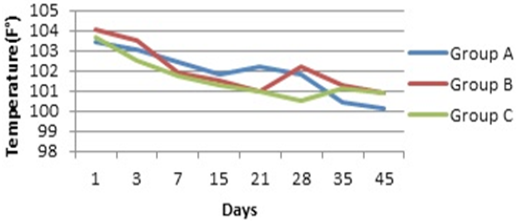
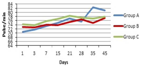
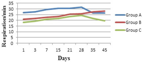
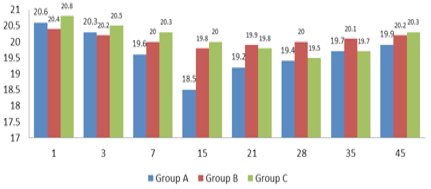
Temperature, pulse and respiration were checked to evaluate general health status which indicated insignificant difference between groups. Graphical presentation of these parameters coincided with (Maish et al., 2005). Various hematological parameters were analyzed to determine effects of esophageal resection on overall body health. There was significant difference between groups in terms of WBCs, RBCs, Hb and ESR. The level of white blood cells elevated initially in group A and B and then gradually decline till end of experiment, whereas least change was observed in group C as compared to group A and B. RBCs and Hb values were at slightly reduced level then normal in group A and B which revealed slight blood loss, reduced local nutritional and oxygen supply post-surgery, while in group C values were in normal range and consistent throughout the experiment. Values of ESR were highly elevated in group A, less in group B and least in group C which indicated presence of high levels of infections and inflammatory changes respectively. These hematological parameters were agreed with that denoted by (Chao et al., 2010) and (Ranen et al., 2004).
Post-surgery, animals were kept off-feed and water for first five days and nutritional supplementation was given by IV line. After 5th day liquid diet was started which was gradually replaced with semisolid and later with solid one with progression of time. Liquid and food intake was least initially in all groups which progressively improved with time. In group wise, overall it was noted that there was minor food and liquid intake in group A, minor to normal in group B and normal in group C from initiation of feed to end of experiment. These results are coincided with that described by (Viklund et al., 2006).
Clinical evaluation of esophageal perforation/stenosis was done by evaluating the frequency of vomiting, subcutaneous emphysema and swallowing difficulty. In average these signs were overall severe in group A, moderate in group B and least in group C which coincided with the findings of (Viklund et al., 2006). Gain/loss in body weight revealed that there was significant difference between groups. Group C showed slight better weight gain till end of experiment whereas no weight gain was observed in group A and B.
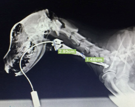
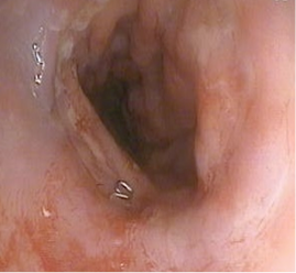
Contrast radiography was used to evaluate luminal diameter/stenosis and leakage at surgical site which revealed that there was significant difference in luminal diameter between groups. Marked decrease in diameter was observed in group A, moderate in group B, while least in group C. These results were similar to that reported by (Serhal et al., 2010) and (Boone et al., 2008). As far as leakage is concerned, radiography revealed that leakage in group A outnumbered group B, while no leakage was seen in group C. These findings were agreed with findings of (Bladergroen et al., 1986). Endoscopy was used to observe level of wound healing and perforations. Wound healing at different experimental intervals revealed that in group A healing was poor, in group B it was moderate to good and in group C it was good to excellent. These findings were agreed with (Hsu et al., 2004). The perforation/leakage evaluation showed that number of perforations was marked in group A, less in group B and none in group C. These endoscopic findings were similar as described by (Hogan et al., 2008).
It was concluded that stapling technique (intra luminal circular stapler 28 mm) has been considered much better then the other two techniques (single layer suturing and double layer suturing). The verdict base explanation for better and reliable result in esophageal anastomosis is that the stapling technique has good results in term of post-op complication, less operative time, give more strength to the anastomosed ends of esophagus, good healing and minimum stenosis. According to our findings double layer suturing technique may be considered as second option due to minimum chances of perforation along with mild fibrosis and stenosis with good healing time. Single layer suturing technique may be third one and least accepted because it experiences more post-op complications, greater chances of infection, dehiscence of anastomosed ends due to less strength and increased stenosis levels.