
Although mounting evidence has shown that mycotoxins can cause intestinal dysfunction and mucosal immune disorders, the effect of zearalenone (ZEA) on intestinal mucosal function remains controversial. We aimed to explore the effects of short-term ZEA exposure on the mucosal barrier function in the colon. We found that the morphology of colonic mucosa in mice was normal after ZEA was administered by gavage for one week, the mRNA expression levels of mucosal Mucin-1, Mucin-2, regenerating islet-derived protein 3 gamma (Reg3γ), and tumor necrosis factor (TNF) were significantly downregulated. The mRNA expression levels of mucosal β-defensin, regenerating islet-derived protein 3 alpha (Reg3a), regenerating islet-derived protein 3 beta (Reg3β) and secretory immunoglobulin A (sIgA) levels were significantly increased. The interleukin-1beta (IL-1β) level was decreased. 16s RNA sequencing further indicated that the colonic microflora had changed, especially the Lactobacillus had increased significantly. Our study showed that short-term ZEA exposure led to resistive increase of colonic probiotics in mice, especially the increase of lactobacillus can reduce intestinal mucosal immunotoxicity.
Research Article · Volume 52, Issue 4, 21 April 2020 · Open Access
Changes of Colon Flora and Antitoxic Effect Induced by Zearalenone in Mice
· · · · · · · · · ·
Affiliations & Notes ⌄
- 1Key Laboratory of Zoonosis Research, Ministry of Education, Jilin University, Changchun 130062, China 2College of Animal Science, Jilin University, Changchun 130062, China Hengtong Fang and Hao Yu contributed equally to this article. , * Corresponding author: yu_hao@jlu.edu.cn
Article Info ⌄
- Journal:
- Pakistan Journal of Zoology
- Published:
- 21 April 2020

From the article
- Further studies found that ZEA exposure significantly reduced the mRNA expression of Mucin 1, Mucin 2 and Reg3γ in colonic mucosa.
- Our study found that ZEA enhanced mucosal immunity in the colon and decreased the colonic Proteobacteria.
- It is worth noting that the intestinal Lactobacillus was increased.
- We believe that intestinal Lactobacillus play an essential role in the intestinal mucosal immune defense against environmental toxins such as mycotoxins.
Selected from the article's own text.
Abstract
INTRODUCTION
According to previous reports, zearalenone (ZEA) is one of the most threatening environmental toxins in the world and it was associated with many fungal diseases in farm animals . Nowadays, we often concern that ZEA induces reproductive toxicity in mammals. There are few studies on the toxic effects of ZEA on intestinal mucosal barriers in animal or human. Studies have revealed that ZEA impairs the small intestinal mucosal barrier , and we have previously demonstrated that ZEA not only disrupts the jejunal microecological balance but also causes inflammation of the jejunal mucosa . But we cannot ignore that colon contains large number of intestinal microbes and its microecological stability is more vulnerable to interference from external substances. Increasing evidence suggests that the incidence of colitis and colon cancer is closely related to environmental pollutants, especially mycotoxins ; ; . Therefore, it is necessary to investigate the effects of ZEA exposure on the colonic mucosal barrier and microecological stability. As we all know, the intestinal mucosa is the first line of defense of the intestinal barrier. Notably, the intestinal epithelial cells, intestinal flora, mucus, sIgA and antimicrobial peptides (AMPs) in the intestinal mucosa constitute the “firewalls” of intestinal mucosal immunity, which protect the body from pathogenic bacteria, virus and environmental pollutants . Recent studies have shown that most mycotoxins can induce intestinal epithelial cell inflammation by destroying intestinal mucosal barrier . Indeed, The intestinal flora plays a role in regulating intestinal immunity, and it acts as an adjuvant to the entire immune system ; . However, there are few studies on the toxic effects of ZEA on the intestinal microflora and colonic mucosal barrier in mammals, and its mechanism is still unclear .
Therefore, we systematically investigated the effects of ZEA on colonic microorganisms and mucosal barriers in mice by 16S rRNA sequencing technology, and explored the potential molecular mechanisms of reduced immunotoxicity, which provided theoretical basis for prevention and treatment of ZEA exposure.
MATERIALS AND METHODS
ZEA (purity > 98%) was purchased from Sigma-Aldrich (St Louis, MO, USA). It was dissolved in ethanol and made into 100 mg/ml. Then the storage solution was dissolved in olive oil at 10mg/ml products before use.
Twenty-four immature male BALB/C mice (Laboratory Animal Center of Jilin University, China), were individually divided into two groups, each group had three replicates, each containing four mice. After a weekend for a trial period, all mice were reared under standard test conditions, and were fed by basal diet (Liaoning Changsheng Biological Company) and sterile drinking water. The recommended dose of ZEA induces a physiological disorder in mice of 20 mg/(kg.bw) (~1% LD50) . We adopted this recommended dose and conducted many tests on mice, we found it was indeed effective. Therefore, the experimental group (ZEA group) was administered ZEA by oral gavage at a dosage of 20 mg/kg body weight (~1% LD50) once daily for one week, and the control group (CON group) was given an equal volume of vector. All experiments were approved by the Institutional Animal Care and Use Committee of Jilin University.
At the end of the experiment, the mice were sacrificed by cervical dislocation. The colon was separated, and the intermediate segment (1-2 cm long) was taken and washed away with saline, and the tissue was fixed with 4% polyoxymethylene. The remaining colon and feces were carefully collected, quickly placed in liquid nitrogen, and then transferred to -80°C.
Intestinal mucosal morphology analysis
The intestinal tissues were fixed with 4% polyoxymethylene and embedded in petrolin, then sectioned into 2-3 µm sections. The sections were stained with hematoxylin first, counterstained with eosin, and finally observed under an optical microscope.
Composition and diversity of the bacterial communities
Total genomic bacterial DNA was extracted from the feces of colon in BALB/c mice. Then the qualified genomic DNA was used for 16S-rDNA V3-V4 region PCR amplification. The primer was 341F (5’ ACTCCTACGGGAGGCAGCAG-3’) and 806R (5’ GGACTACHVGGGTWTCTAAT-3’). All the procedures were performed according to the manufacturer’s protocol (BGI, Shengzhen, China). Two hypervariable regions of 16S rDNA, the V3 and V4 regions, were used to identify the vast majority of the bacteria based on the 16S rDNA sequencing. The qualified PCR products were purified by 16S V3-V4 amplification, and the DNA library was constructed. The qualified DNA library was sequenced using an Illumina MiSeq 2*300, and information was acquired for the bioinformatic analysis.
The raw data were filtered to eliminate adapter pollution and low quality to obtain clean reads. Then, the paired-end reads with overlapping regions were merged to raw tags with FLASH software (v1.2.11) . Paired-end reads without overlaps were removed. Following removal of the primer sequences, the forward and reverse amplification primers were mapped to the two ends of the tags. The tags were clustered to the operational taxonomic unit (OUT) at 97% sequence similarity by scripts using the software USEARCH (v7.0.1090) (RC, 2013). The PCR-generated chimaera from the OTU representation sequence was removed, and effective tags were obtained by UCHIME software (v4.2.40) . The database used for species annotation was Greengene (V201305) .
Determination of the relative expression of mRNA in intestinal mucosal barrie by real-time PCR
Total RNA was extracted from colon using TriPure (Roche, Basel, Switzerland) and the cDNA was synthesized using Reverse Transcription System (Promega, Madison, USA) and SYBR Green Mix Kit (Trans, Beijing, China) was used for real-time PCR. Primer design as shown in Supplementary Table I.
Detection of sIgA in the colon
Equal amounts of colonic feces were dissolved with 0.01 M PBS, and broken down at low temperature, centrifuged for 10 min at 3000 r, then the supernatant was collected. The total protein concentration of the feces was detected by BCA. Then the absorbance was detected at 562 nm according to the instructions of mouse sIgA ELISA kit (eBioscience, California, USA), and the sIgA content of feces was calculated (ng/ml).
Detection of cytokine in the colon
Colon was homogenized with 0.01 M PBS to make 10% tissue homogenate. Then at low temperature, the homogenate was centrifuged for 10 min at 3000 r. The supernatant was used for estimation of total protein according to the instructions of IL-1β, IL-10, and TNF (Naniing Jiancheng Bioengineering Institute, Nanjing, China). The cytokine levels were calculated as ng/g prot.
Statistical analysis
All the data were analysed using SPSS statistical software (version 20.0). The variation between the CON group and the ZEA group was analysed by a t-test, and the results are expressed as the mean ± SEM. The differences were judged as statistically significant at P values < 0.05.
RESULTS
Effect of ZEA on mucosal morphology
To objectively assess the effects of ZEA on intestinal mucosa, the histological sections of colon mucosal morphology was observed, as shown in Figures 1A, 1B. We found that there was no significant pathological change between ZEA group and control group, but the intestinal cavity of the ZEA group became significantly larger (Fig. 1C).
Table I. Effect of ZEA on bacterial community diversity in the colonic digesta.
| Sample name | Tag number | OTU number | Length(bp) | ace | simpson |
| CON | 25379 | 286 | 415 | 304.4 | 0.057 |
| ZEA | 24024 | 295 | 416 | 318.0 | 0.035 |
ZEA changes the balance of the intestinal flora
In order to study the effect of ZEA on intestine microflora in the mice, a total of 148209 effective tags were obtained by 16S-rRNA sequencing, with an average of 24702 effective tags for each sample and an average length of 416 bp (Table I). Using the ACE index (Table I) to assess bacterial richness, short-term ZEA exposure led to a tendency to increase the richness of the colonic microflora. At the same time, the Simpson index was used to evaluate changes in the intestinal microbial diversity (Table I). In the ZEA group, the bacterial diversity was almost similar to that of the CON group, but the diversity of the colonic microflora showed a tendency to increase. At 97% similarity, the colonic bacterial community of the CON and ZEA groups shared 325 OTUs (Fig. 2A). The flattening of the rarefaction curve based on the values of observed species (Fig. 2B) indicated that our data volume covered all species of the community in the colon samples. At phylum level (Fig. 2C), compared with the CON group, the abundance of Proteobacteria in the ZEA group was reduced more than 2-fold (P < 0.001). The abundance of Actinobacteria in the ZEA group was increased (P < 0.01). At genus levels (Fig. 2C), Lachnoclostridium and Alloprevotella were highly abundant. Compared to the CON group, ZEA reduced Lachnoclostridium from 16.7% to 9.8% (P > 0.05) and reduced Alloprevotella from 9.6% to 4.9% (P > 0.05). The abundance of Desulfovibrio was reduced from 3.1% to 1.5% (P > 0.05). Compared with CON group, the abundance of Anaeroplasma, Flavonifractor, Helicobacter, Prevotella and Ruminiclostridium in the ZEA group was decreased (P > 0.05). Interestingly, the Lactobacillus was upregulated more than 4-fold (P < 0.05). In addition, we found that short-term ZEA exposure led the emergence of Akkermansia in colon.
Effect of ZEA on intestinal mucosal barrier in mice
We further detected the effect of ZEA on colonic mucosal barrier. We examined the mRNA expression of key genes that play the first line of defense in the colonic mucosa (Fig. 3). The mRNA expression of Mucin 1, Mucin 2, and Reg3γ were significantly downregulated by ZEA exposure. Interestingly, ZEA increased mRNA expression of β-defensin, Reg3α, and Reg3β. In addition, the mRNA expression of inflammatory cytokines IL-1β and TNF were remarkably downregulated after ZEA exposure. There was no significant change in the mRNA level of IL-10.
The effect of ZEA on intestinal mucosal immunity
To assess the effect of ZEA on intestinal mucosal immunity, we firstly detect the faecal sIgA levels in the colon. The results showed that short-term ZEA exposure induced the mucosal sIgA (P < 0.001) and the faecal IgA in the ZEA group increased more than 3-fold compared to that in the CON group (Fig. 4A). At the same time, we investigated the effects of ZEA on cytokine secretion involved in the inflammatory response in the colon. Interestingly, ZEA inhibited the secretion of IL-1β in the colon, but there was no change in TNF and IL-10 levels (Fig. 4B).
DISCUSSION
Mycotoxin is a class of environmental pollutant that easily causes an intestinal ecological imbalance and induces some intestinal diseases ; ; . We found that the structure of colonic microbial communities was altered by short-term ZEA exposure. Changes in some of the dominant bacteria in the gut indicated that our ZEA exposure induced disturbances in the intestinal microecology. We found that Proteobacteria which are associated with inflammation was decreased . At the same time, Lachnoclostridium and Alloprevotella, Desulfovibrio,
Anaeroplasma, Flavonifractor, Helicobacter, Prevotella and Ruminiclostridium were decreased. Previous studies have shown that Clostridium leptum, Lachnospiraceae, and Prevotellaceae were reduced in inflammatory bowel disease patients . Changes in these micrbiotes suggest instability of the intestinal flora or sensitivity of the intestines. It is worth noting that ZEA increased the Lactobacillus. Piotrowska et al. (2014) argued that long-term exposure to low doses of ZEA caused an increase in the Lactobacillus in the colon, but the change was not significant. Reddy et al. (2018) found that the ZEA-contaminated feed significantly affected the colon microbiota, especially Lactobacillus, which was increased by 2.7%. They speculate that Lactobacillus plays a major role in detoxification of these mycotoxins. Indeed, Lactobacillus have been evaluated in studies in animals and humans with respect to antibiotic-associated diarrhoea, travellers’ diarrhoea, pediatric diarrhoea, inflammatory bowel disease, and irritable bowel syndrome. Lactobacillus is not only beneficial to intestinal health but also combined with mycotoxins to remove mycotoxins . The decrease of Proteobacteria and the increase of Lactobacillus seem to indicate that damage to the colon by ZEA has been controlled, as evidenced by normal colon morphology. We suspect that the increase of the intestinal cavity area may also be due to the changes in intestinal flora.
Further studies found that ZEA exposure significantly reduced the mRNA expression of Mucin 1, Mucin 2 and Reg3γ in colonic mucosa. Current studies suggest that ZEA increases the secretion of mucin in Caco-2/HT29-MTX cells . Reg3γ and mucins are used to prevent microbial contact with the epithelial layer ; ; . The decrease of these mRNA expressions indicates the decrease of mucosal defense. Interestingly, ZEA increased the mRNA expression of colonic mucosal β-defensin, Reg3α and Reg3β. In addition, the sIgA in the colon was also increased. Indeed, β-defensin Reg3α/β and sIgA play an essential role in the immune defense of the intestinal mucosa .
ZEA has been described as either an inductor or a suppressor of pro-inflammatory cytokines ; . Our study found that ZEA exposure inhibited the IL-1β and the mRNA expression of TNF was downregulated. Fan et al. (2017) suggested that ZEA leads to increased levels of IL-1β in colon tissue of mice. It contradicts our findings. However, Liu et al. (2014) revealed that different doses of ZEA showed a downward trend of IL-1β and TNF in the jejunum of rats. Braicu et al. (2016) found that exposure to ZEA caused a decrease in the IL-1β and TNF in animal which is still waiting for us to further study.
Current study suggests that ZEA is absorbed through the small intestine and some of it is degraded through the circulation in the liver and intestine while some combines with intestinal microbes. Eventually, only a small amount of ZEA and its degradation products act on the large intestine . On one hand, a low dose of ZEA is considered to be immunosuppressant and may inhibit the inflammatory response. On the other hand, the increase of colonic Lactobacillus only remove mycotoxins by combining with mycotoxins but also regulate intestinal mucosal immunity by producing short chain fatty acid (SCFA) ; . Thereby mucosal damage is alleviated to some extent. But the specific reasons are still waiting to be explored.
CONCLUSION
Our study found that ZEA enhanced mucosal immunity in the colon and decreased the colonic Proteobacteria. It is worth noting that the intestinal Lactobacillus was increased. We believe that low doses of ZEA enter the colon and stimulate the growth of beneficial bacteria in the colon (Fig. 5), which indirectly activates the mucosal defense system. Ultimately the toxicity of ZEA to the intestinal mucosa was alleviated. All of this credit comes from the special role of Lactobacillus, which play a key role in regulating intestinal mucosal immunity and promoting colon health. Thus, the colonic mucosa can protect against a certain degree of toxins such as ZEA. This appears to provide us with a new way of thinking about controlling mycotoxins. We believe that intestinal Lactobacillus play an essential role in the intestinal mucosal immune defense against environmental toxins such as mycotoxins.
ACKNOWLEDGEMENTS
This study was financially supported by the Jilin Scientific and Technological Development Program (20170307018NY, 20190301034NY); the Special Project of the Province-University Co-constructing Program of Jilin Province (SXGJXX2017-4); the central finance forestry science and technology promotion demonstration fund project and the National Key Research and Development Program of China Stem Cell and Translational Research (2017YFA0105101); the Open Projects of Northeast Agricultural University/Scientific Observing and Experimental Station of Animal Nutrition and Feed Science in Northeast, Ministry of Agriculture, P.R. China; and the Jilin Modern Agricultural Technology Demonstration and Extension Project.
Supplementary material
There is supplementary material associated with this article. Access the material online at: https://dx.doi.org/10.17582/journal.pjz/20190115060153
Statement of conflict of interest
The authors declare that there are no conflicts of interest.
Figures
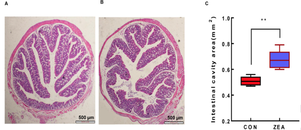
The effect of ZEA on the intestinal mucosal morphology of the colon in BALB/C mice. A: The colon of the CON group. B: The colon of the ZEA group. C: The area of intestinal cavity. **: P value<0.01.
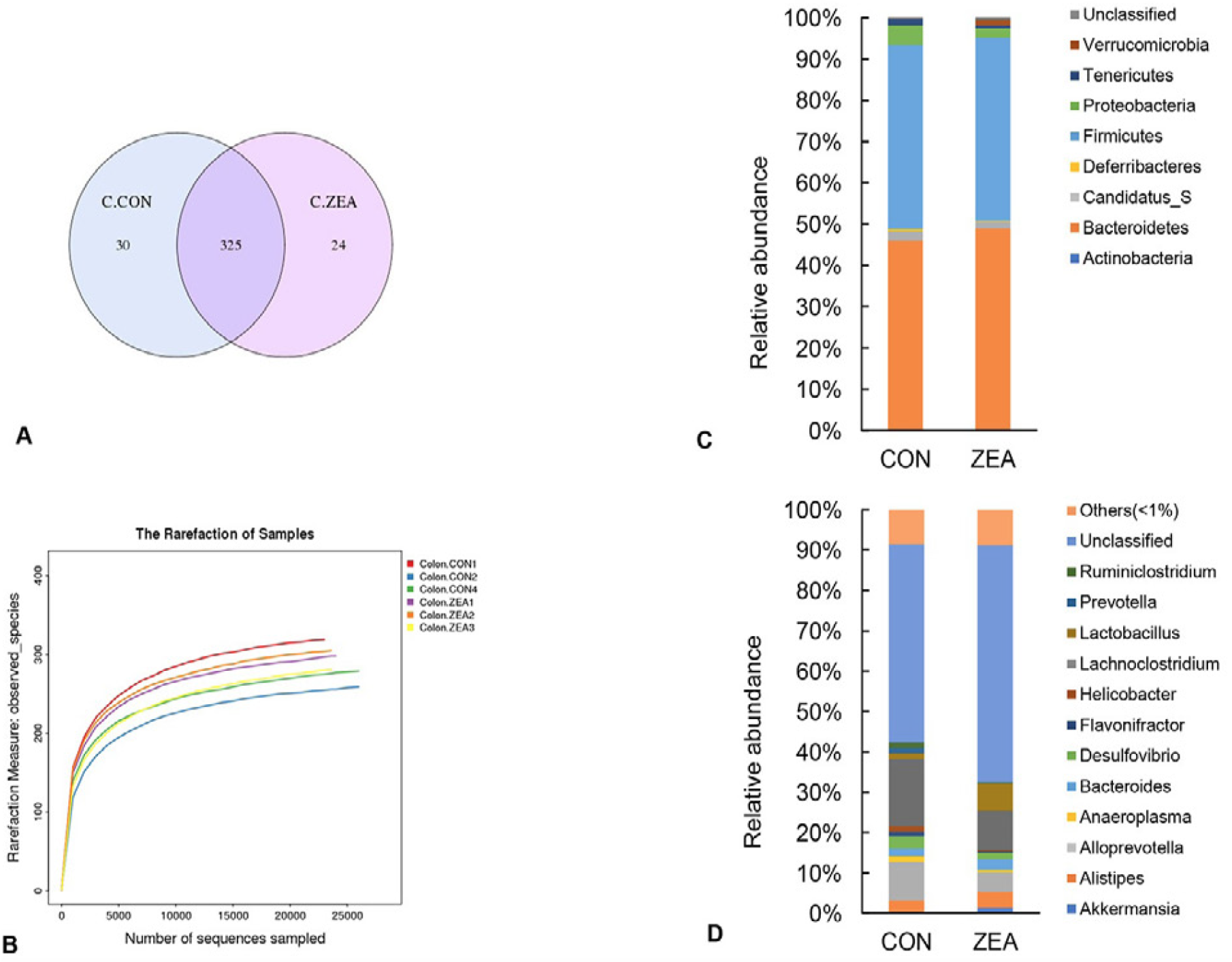
Effect of ZEA on the intestinal microflora. (A), comparison of OTU. (B), rarefaction curves based on the observed species values. (C), relative abundance of different bacterial phylum. (D), relative abundance of different bacterial genus.
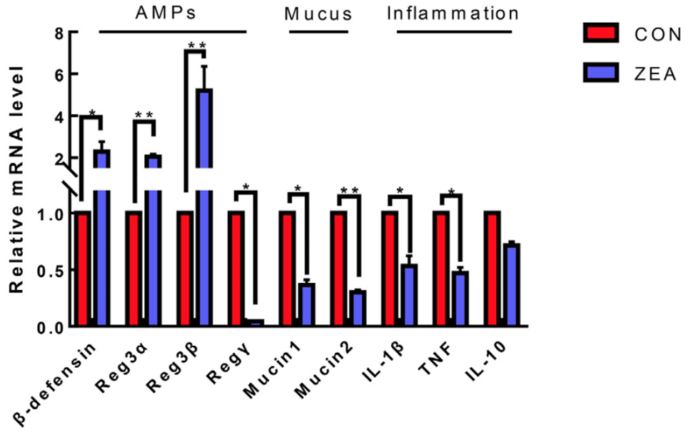
Changes in the immune gene expression in the colonic mucosa of the mice. All the data are expressed as the means ± SEM. *: P value<0.05, **: P value<0.01.
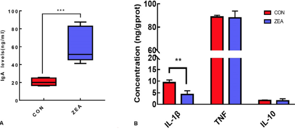
(A), Changes in the sIgA levels in the colonic faeces. (B), Changes in the cytokines in the colon. All the data are expressed as the means ± SEM. **: P value<0.01, ***: P value<0.001.
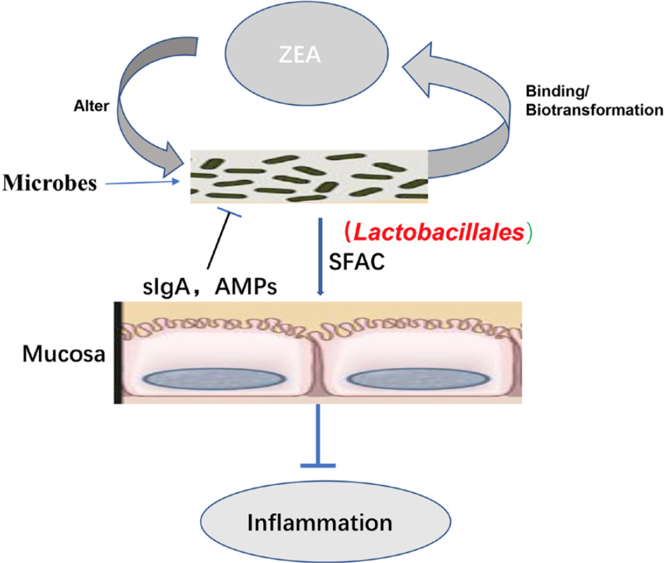
Schematic diagram of ZEA acting on colonic mucosa.
References
- Belkaid, Y. and Hand. T.W., 2014. Role of the microbiota in immunity and inflammation. Cell, 157: 121-141. https://doi.org/10.1016/j.cell.2014.03.011[CrossRef]
- Bondy, G.S. and Pestka. J.J., 2000. Immunomodulation by fungal toxins. J. Toxicol. environ. Hlth. B. Crit. Rev., 3: 109-143. https://doi.org/10.1080/109374000281113[CrossRef]
- Braicu, C., Cojocneanupetric, R., Jurj, A., Gulei, D., Taranu, I., Gras, A.M., Marin, D.E. and Berindanneagoe, I., 2016. Microarray based gene expression analysis of sus scrofa duodenum exposed to zearalenone: significance to human health. Bmc Genom., 17:646. https://doi.org/10.1186/s12864-016-2984-8[CrossRef]
- Carvalho, F.A., Koren, O., Goodrich, J.K., Johansson, M.E.V., Nalbantoglu, I., Aitken, J.D., Su, Y., Chassaing, B., Walters, W.A. and González. A., 2012. Transient inability to manage proteobacteria promotes chronic gut inflammation in TLR5-deficient mice. Cell Host Microbe, 12: 139-152. https://doi.org/10.1016/j.chom.2012.07.004[CrossRef]
- Choung, R.S. and Iii, G.R.L., 2011. Epidemiology of IBS. Gastroenterol. Clin. N., 40: 1-10. https://doi.org/10.1016/j.gtc.2010.12.006[CrossRef]
- Compare, D., and Nardone, G., 2014. The role of gut microbiota in the pathogenesis and management of allergic diseases. Eur. Rev. Med. Pharmacol. Sci., 18: 11-17.
- Desantis, T. Z., Hugenholtz, P., Larsen, N., Rojas, M., Brodie, E.L., Keller, K., Huber, T., Dalevi, D., Hu, P. and Andersen, G.L., 2006. Greengenes, a chimera-checked 16S rRNA gene database and workbench compatible with ARB. Appl. environ. Microbiol., 72: 5069-5072. https://doi.org/10.1128/AEM.03006-05[CrossRef]
- Edgar, R. C., Haas, B. J., Clemente, J. C., Christopher, Q. and Rob, K., 2011. UCHIME improves sensitivity and speed of chimera detection. Bioinformatics, 27: 2194-2200. https://doi.org/10.1093/bioinformatics/btr381[CrossRef]
- Edgar, R.C., 2013. UPARSE: highly accurate OTU sequences from microbial amplicon reads. Nat. Methods, 10: 996-998. https://doi.org/10.1038/nmeth.2604[CrossRef]
- Elnezami, H.S., Chrevatidis, A., Auriola, S., Salminen, S. and Mykkänen, H., 2002. Removal of common Fusarium toxins in vitro by strains of Lactobacillus and Propionibacterium. Fd. Addit. Contam., 19: 680-686. https://doi.org/10.1080/02652030210134236[CrossRef]
- Fan, P., Song, P., Li, L., Chang, H., Chen, J., Yang, W., Qiao, S., Wu, G., Zhang, G. and Xi, M., 2017. Roles of biogenic amines in intestinal signaling. Curr. Protein Pept. Sci., 18: 532-540. https://doi.org/10.2174/1389203717666160627073048[CrossRef]
- Fan, W., Lv, Y., Ren, S., Shao, M., Shen, T., Huang, K., Zhou, J., Yan, L. and Song. S., 2017b. Zearalenone (ZEA)-induced intestinal inflammation is mediated by the NLRP3 inflammasome. Chemosphere, 190: 272-279. https://doi.org/10.1016/j.chemosphere.2017.09.145[CrossRef]
- Fink-Gremmels, J. and Malekinejad. H., 2007. Clinical effects and biochemical mechanisms associated with exposure to the mycoestrogen zearalenone. Anim. Feed Sci. Tech., 137: 326-341. https://doi.org/10.1016/j.anifeedsci.2007.06.008[CrossRef]
- Gajęcka, M., Zielonka, Ł. and Gajęcki. M., 2016. Activity of zearalenone in the porcine intestinal tract. Molecules, 22: 18. https://doi.org/10.3390/molecules22010018[CrossRef]
- Kollarczik, B., Gareis, M. and Hanelt, M., 2010. In vitro transformation of the fusarium mycotoxins deoxynivalenol and zearalenone by the normal gut microflora of pigs. Nat. Toxins, 2: 105-110. https://doi.org/10.1002/nt.2620020303[CrossRef]
- Liu, M., Gao, R., Meng, Q., Zhang, Y., Bi, C. and Shan. A., 2014. Toxic effects of maternal zearalenone exposure on intestinal oxidative stress, barrier function, immunological and morphological changes in rats. PLoS One, 9: e106412. https://doi.org/10.1371/journal.pone.0106412[CrossRef]
- Macpherson, A.J., Slack, E., Geuking, M.B. and Mccoy, K.D., 2009. The mucosal firewalls against commensal intestinal microbes. Semin. Immunopathol., 31: 145-149. https://doi.org/10.1007/s00281-009-0174-3[CrossRef]
- Magoč, T. and Salzberg, S.L., 2011. FLASH: fast length adjustment of short reads to improve genome assemblies. Bioinformatics, 27: 2957-2963. https://doi.org/10.1093/bioinformatics/btr507[CrossRef]
- Maresca, M. and Fantini, J., 2010. Some food-associated mycotoxins as potential risk factors in humans predisposed to chronic intestinal inflammatory diseases. Toxicon, 56: 282-294. https://doi.org/10.1016/j.toxicon.2010.04.016[CrossRef]
- Marin, D.E., Taranu, I., Burlacu, R., Manda, G., Motiu, M., Neagoe, I., Dragomir, C., Stancu, M. and Calin. L., 2011. Effects of zearalenone and its derivatives on porcine immune response. Toxicology in vitro., 25: 1981-1988. https://doi.org/10.1016/j.tiv.2011.06.022[CrossRef]
- Martín, R., Miquel, S., Ulmer, J., Kechaou, N., Langella, P., and Bermúdezhumarán, L.G., 2013. Role of commensal and probiotic bacteria in human health: a focus on inflammatory bowel disease. Microbe Cell Fact., 12: 71-71. https://doi.org/10.1186/1475-2859-12-71[CrossRef]
- Molloy, M.J., Bouladoux, N. and Belkaid, Y., 2012. Intestinal microbiota: Shaping local and systemic immune responses. Semin. Immunol., 24: 58-66. https://doi.org/10.1016/j.smim.2011.11.008[CrossRef]
- Muriel, D., Vlieg, J.E.T.H., 2015. Fate, activity, and impact of ingested bacteria within the human gut microbiota. Trends Microbiol., 23: 354-366. https://doi.org/10.1016/j.tim.2015.03.002[CrossRef]
- Piotrowska, M., Śliżewska, K., Nowak, A., Zielonka, Ł., Żakowska, Z., Gajęcka, M. and Gajęcki, M., 2014. The effect of experimental fusarium mycotoxicosis on microbiota diversity in porcine ascending colon contents. Toxins, 6: 2064-2081. https://doi.org/10.3390/toxins6072064[CrossRef]
- Reddy, K.E., Jeong, J.Y., Song, J., Lee, Y., Lee, H.J., Kim, D.W., Jung, H.J., Kim, K.H., Kim, M., Oh, Y.K., Lee, S.D. and Kim, M., 2018. Colon microbiome of pigs fed diet contaminated with commercial purified deoxynivalenol and zearalenone. Toxins, 10: 347. https://doi.org/10.3390/toxins10090347[CrossRef]
- Richards, J.D., Lange, J.G.F.M.D., 2005. The gastrointestinal microbiota and its role in monogastric nutrition and health with an emphasis on pigs: current understanding, possible modulations, and new technologies for ecological studies. Can. J. Anim. Sci., 85: 421-435. https://doi.org/10.4141/A05-049[CrossRef]
- Salah-Abbès, J.B., Abbès, S., Houas, Z., Abdel-Wahhab, M.A. and Oueslati, R., 2010. Zearalenone induces immunotoxicity in mice: Possible protective effects of radish extract (raphanus sativus). J. Pharm. Pharmacol., 60: 761-770. https://doi.org/10.1211/jpp.60.6.0012[CrossRef]
- Sanchez de Medina, F., Romero-Calvo, I., Mascaraque, C. and Martinez-Augustin. O., 2014. Intestinal inflammation and mucosal barrier function. Inflamm. Bowel Dis., 20: 2394-2404. https://doi.org/10.1097/MIB.0000000000000204[CrossRef]
- Seonghwan, P., Hyejin, C., Yang, H., Keehun, D., Juil, K. and Yuseok, M., 2010. Repression of peroxisome proliferator-activated receptor γ by mucosal ribotoxic insult-activated CCAAT/enhancer-binding protein homologous protein. J. Immunol., 185: 5522-5530. https://doi.org/10.4049/jimmunol.1001315[CrossRef]
- Sonnenburg, Justin, L., Xu, J., Leip, D., Chen, C.-H., Westover, B.P., Weatherford, J., Buhler, J.D. and Gordon, J.I., 2005. Glycan foraging in vivo by an intestine-adapted bacterial symbiont. Science, 307: 1955-1959. https://doi.org/10.1126/science.1109051[CrossRef]
- Stefania, D.S., Elisabetta, C., Mauro, M., Emilio, J. and Marcello, C., 2015. Nutritional keys for intestinal barrier modulation. Front. Immunol., 6: 612. https://doi.org/10.3389/fimmu.2015.00612[CrossRef]
- Swidsinski, A., Ladhoff, A., Pernthaler, A., Swidsinski, S., Loening–Baucke, V., Ortner, M., Weber, J., Hoffmann, U., Schreiber, S. and Dietel, M., 2002. Mucosal flora in inflammatory bowel disease. Gastroent. Hepat., 122: 44-54. https://doi.org/10.1053/gast.2002.30294[CrossRef]
- Turner, J.R., 2009. Intestinal mucosal barrier function in health and disease. Nat. Rev. Immunol., 9: 799-809. https://doi.org/10.1038/nri2653[CrossRef]
- Wan, L.Y.M., Allen, K.J., Turner, P.C. and Elnezami. H., 2014. Modulation of mucin mRNA (muc5ac and muc5b) expression and protein production and secretion in caco-2/ht29-mtx co-cultures following exposure to individual and combined fusarium mycotoxins. Toxicol. Sci., 139: 83-98. https://doi.org/10.1093/toxsci/kfu019[CrossRef]
- Wang, J., Tang, H., Zhang, C., Zhao, Y., Derrien, M., Rocher, E., van-Hylckama, J., Strissel, K., Zhao, L., Obin, M. and Shen, J., 2015. Modulation of gut microbiota during probiotic-mediated attenuation of metabolic syndrome in high fat diet-fed mice. The ISME J., 9: 1–15. https://doi.org/10.1038/ismej.2014.99[CrossRef]
- Wang, X., Yu, H., Shan, A.S., Jin, Y. C., Fang, H.T., Zhao, Y., Shen, J., Zhou, C., Zhou, Y.F., Fu, Y.R., Wang, J.M. and Zhang. J., 2018. Toxic effects of zearalenone on intestinal microflora and intestinal mucosal immunity in mice. Fd. Agric. Immunol., 29: 1002-1011. https://doi.org/10.1080/09540105.2018.1503233[CrossRef]
- Wild, C.P. and Gong, Y.Y., 2010. Mycotoxins and human disease: a largely ignored global health issue. Carcinogenesis, 31: 71-82. https://doi.org/10.1093/carcin/bgp264[CrossRef]
- Zinedine, A., Soriano, J.M., Moltó, J.C., and Manes. J., 2007. Review on the toxicity, occurrence, metabolism, detoxification, regulations and intake of zearalenone: an oestrogenic mycotoxin. Fd. Chem. Toxicol., 45: 1-18. https://doi.org/10.1016/j.fct.2006.07.030[CrossRef]
Supplementary material
Services
Article usage statistics
Loading…