Wounds break the continuity of the skin and allow organisms to gain access to tissues and cause infection, particularly those that inoculate pathogens into the dermis, frequently cause skin infections. The main cause of open wounds is physical trauma or injury while fighting or bite by another animal, during transportation accidents or due to saddle misfit injuries (Figure 4 and 5). In present study out of 255, 9 samples were rendered negative as no microbial growth appeared till 76 hrs., while 246 samples revealed presence of different species of bacteria and in few cases other agents as fungi (Table 1 and 2). As present study mainly focused on bacterial etiology, other (fungal) agents are not being discussed here, out of 255, 9(3.53%) samples of wounds were negative and 246(96.47%) were positive. Microbiological investigation based upon cultural, morphological and biochemical studies revealed isolation of strains of Staphylococcus aureus, Pseudomonas aeruginosa,
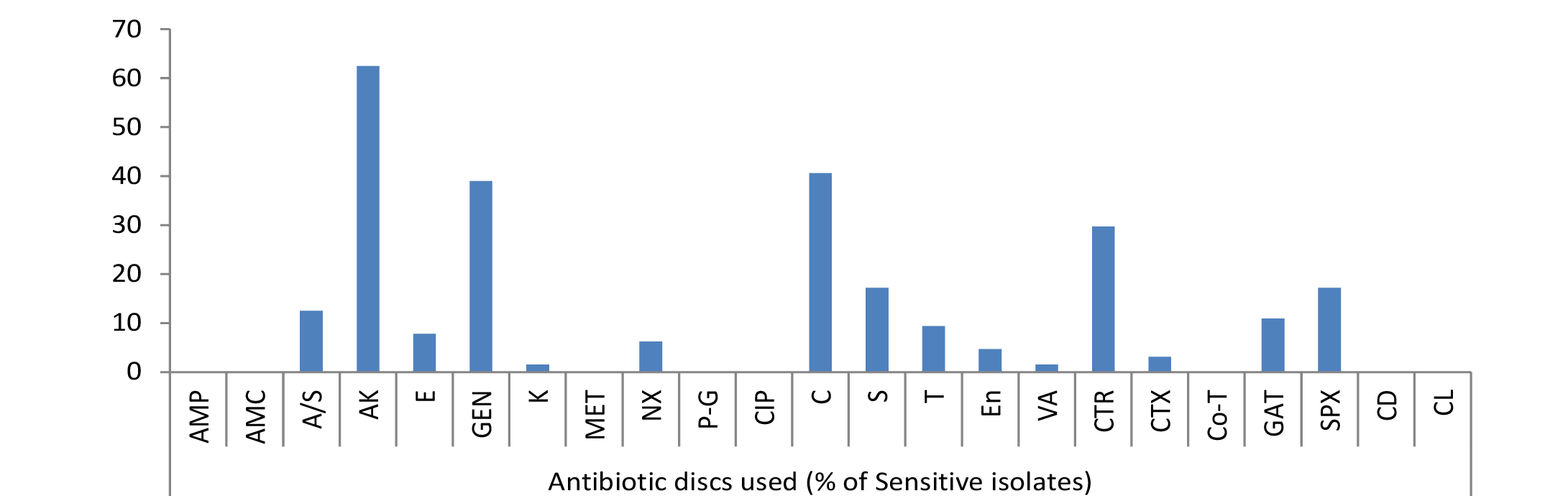
Figure 1: Percentage sensitivity of E. coli isolates
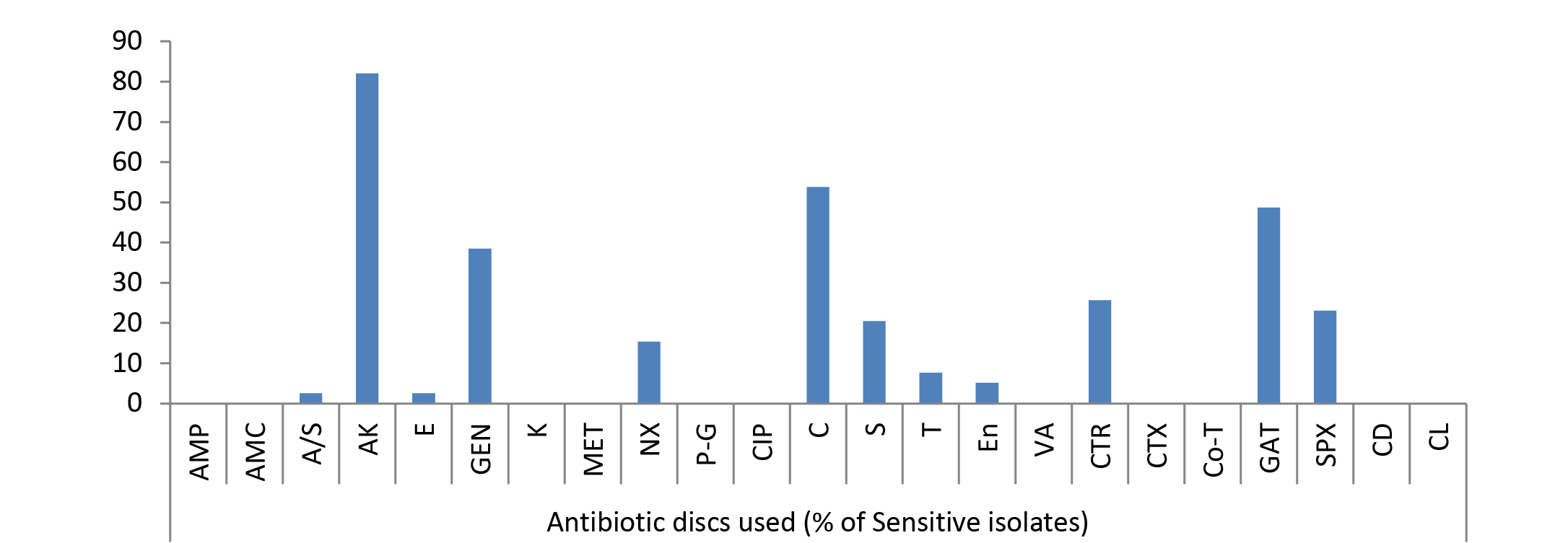
Figure 2: Percentage sensitivity of Pseudomonas spp. isolates
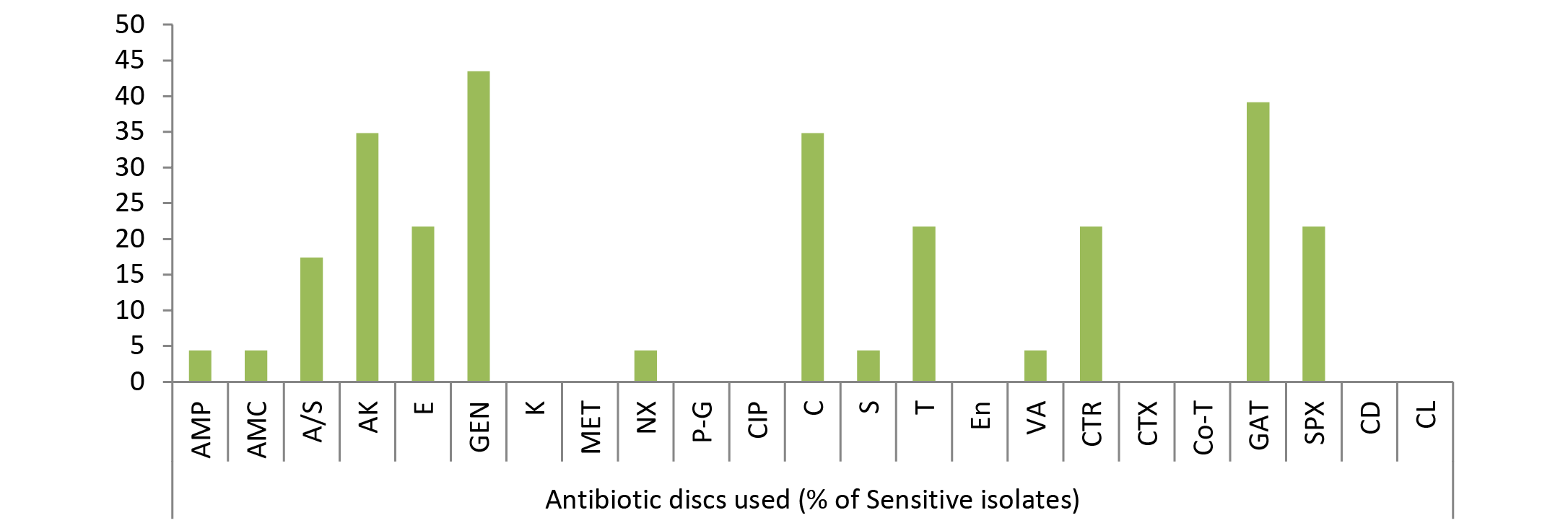
Figure 3: Percentage sensitivity of Klebsiella spp. isolates
E.coli, Klebsiella spp., Protius, Bacillus, Clostridium, Streptococcus, Micrococcus etc. (Figure 6). The majority of wounds contained a mixture of aerobes and anaerobes, which reflect the diversity of microorganisms present in the atmosphere infecting wounds and vicinity of the affected animal and to a lesser extent of their skin also.
Table 3: Result of antibiotic sensitivity testing of various isolates recovered from wounds of animals
AMP= Ampicillin; AMC= Amoxycillin; A/S= Ampicillin/Sulbactum; AK= Amikacin; E= Erythromycin; CTX= Cefotaxime; GEN= Gentamicin; CIP= Ciprofloxacin; K= Kanamycin; MET= Methicillin; T = Tetracycline; Nx= Norfloxacin; P-G= Penicillin-G; C= Chloramphenicol; S= Streptomycin; En= Enrofloxaacin; Va= Vancomycin; CTR= Ceftriaxone; Co-T= Cotrimoxazole; GAT= Gatifloxacin; SPX= Sparfloxacin; CD= Clindamycin; CL= Colistin
The results of frequency distribution of bacterial isolates were presented in (Table 2). In the study performed over animals for wounds among significant isolates Staphylococcus aureus was the most common isolate (36.22 %), followed by E. coli (34.59 %), Pseudomonas (20.54), Bacillus (16.21) and Klebsiella (12.43). Other bacterial agents involved were less than 10 % as of Micrococcus spp (8.11 %), Streptococcus pyogens (7.56), Proteus (6.49), Clostridium (3.78), Gram negative non-lactose fermenter (2.7%), Gram positive non-spore producing bacilli (2.16) and Fusobacterium (0.54 %). Clostridium spp. and Fusobacterium were the only obligate anaerobes isolated from the samples collected. Bacterial skin infections common in animal veterinary practice, are most frequently caused by Staphylococcus aureus apart from other common causative agents like Escherichia coli as evident from the results. Gram negative bacteria are generally secondary invaders which establishes under chronic conditions. Pseudomonas, however, is a Gram negative notorious bacterium while involvement of Bacillus in 16.21% cases indicated that it is actively causing wound infection rather than present as contaminant because samples were collected under aseptic conditions. Single isolate of Fusobacterium was also identified; one probable reason behind this low prevalence of this microorganism could be good managemental condition on the farm for animal rearing or slow growing tendency of Fusobacterium.
Due to the scarcity of literature on animal studies in this regard present experiment was designed to look in to the pattern of bacterial susceptibility of wound isolates against various antimicrobial drugs. The in vitro antibiotic sensitivity test of different types of all bacterial isolates to 23 different antibiotics such as Ampicillin, Amoxycillin, Ampicillin/Sulbactum, Amikacin, Erythromycin, Gentamicin, Kanamycin, Methicillin, Norfloxacin, Penicillin-G, Ciprofloxacin, Chloramphenicol, Streptomycin, Tetracycline, Enrofloxacin, Vancomycin, Ceftriaxone, Cefotaxime, Cotrimoxazole, Gatifloxacine, Sparfloxacine, Clindamycin and Colistin was studied. A slight variation was noticed in the results of sensitivity of isolates against 23 different antibiotics used, though no single antibiotic therapy was found effective against all the bacteria. Antibiotic sensitivity pattern of most of bacteria, isolated from different cases of skin/wound infections, revealed resistant strains even against multiple drugs. Gatifloxacin, sparfloxacin, amikacin, chlloramphenicol and gentamicin were highly sensitive drugs in most of the cases (Figure 1, 2 and 3). The isolated bacteria were less sensitive to erythromycin, colistin, cefotaxime, Enrofloxacin, Vancomycin, Tetracycline, Ceftriaxone, Clindamycin, Streptomycin, and Kanamycin while resistant to Ampicillin, Amoxycillin, Ampicillin/Sulbactum, Erythromycin, Methicillin and Norfloxacin in most of cases (Table 3).
Table 4: Sample of wounds over abnormal skin/tissue outgrowth
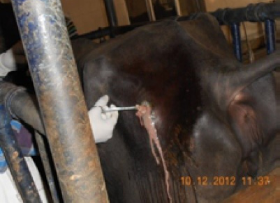
Figure 4: Gun-shot wound in buffalo
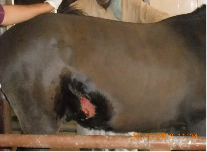
Figure 5: Lacerated wound in horse
Among Streptococcus pyogens 40% isolates against gatifloxacin and 33.3% against amikacin were sensitive, rest 26.67% were mild sensitive to chloramphenicol and tetracycline, 13.33% to erythromycin and vancomycin, 6.67% to ampicillin, norfloxacin and streptomycin, 4.48% to gentamicin, ceftriaxone, sparfloxacin while no strains were sensitive to amoxycillin, ampicillin/sulbactum, kanamycin, methicillin, penicillin-G, ciprofloxacin, enrofloxacin, cefotaxime, cotrimoxazole, clindamycin and colistin.
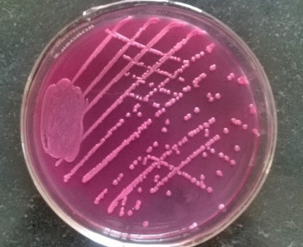
Figure 6: Pink colonies over MLA
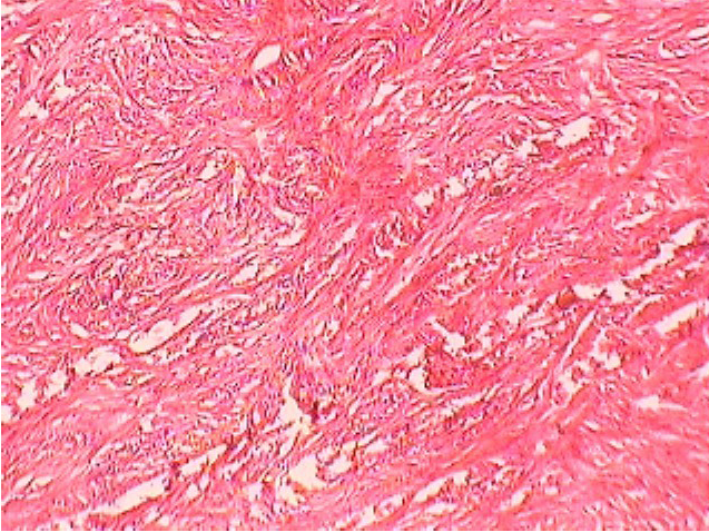
Figure 7: Fibro-sarcoma in Buffalo
Out of 34.59% E. coli isolates, maximum sensitivity was shown by 62.5% strains against amikacin but no other drug showed sensitivity of more than 50%. Among rest isolates 40.62% to chloramphenicol, 39% to gentamicin, 29.69% to ceftriaxone, 17.19% to streptomycin and sparfloxacin, 12.5% to ampicillin/sulbactum, 10.94% to gatifloxacin, 9.38% to tetracycline, 7.81% to erythromycin, 6.25% to norfloxacin, 4.69% to enrofloxacin, 3.12% to cefotaxime, 1.56% to kanamycin and vancomycin were mild sensitive while ampicillin, amoxycillin, methicillin, penicillin-G, ciprofloxacin, cotrimoxazole, clindamycin and colistin were resistant drugs (Figure 1).
Out of 23 isolates of Klebsiella spp., 43.48% were sensitive to gentamicin; 39.13% to gatifloxacin; 34.78% to amikacin, chloramphenicol; 21.74% to sparfloxacin, ceftriaxone, tetracycline, erythromycin; 17.39% to ampicillin/sulbactum, 4.35% to ampicillin, amoxycillin, norfloxacin, streptomycin, vancomycin (Figure 3) while kanamycin, methicillin, penicillin-G, ciprofloxacin, enrofloxacin, cefotaxime, cotrimoxazole, clindamycin and colistin drugs failed to show sensitivity as no zone of inhibition was produced against these antimicrobial discs.
Amongst 39% Pseudomonas spp. isolates recovered from wound cases 82.05% strains showed maximum sensitivity to amikacin followed by 53.84% strains sensitive to chloramphenicol (Figure 2). Rest of drugs showed mild sensitivity, 48.72% isolates sensitive to gatifloxacin; 38.46% to gentamicin; 25.64% to ceftriaxone; 23.07% to sparfloxacin; 20.51% to streptomycin; 15.38% to norfloxacin; 7.69% to tetracycline; 5.13% to enrofloxacin; 2.56% to erythromycin, ampicillin/Sulbactum; and ampicillin, amoxycillin, kanamycin, methicillin, penicillin-G, ciprofloxacin, vancomycin, cefotaxime, cotrimoxazole, clindamycin and colistin were resistant.
As evident from Table 3 result of current study illustrated that among isolated bacteria Staphylococcus aureus, Streptococcus pyogens, Micrococcus and Proteus were found most sensitive against gatifloxacin; E. coli, Bacillus, Pseudomonas, Clostridium against amikacin and Klebsiella against gentamicin. Single isolate of Fusobacterium was sensitive against streptomycin, Gram negative non-lactose fermenters showed maximum sensitivity to amikacin while Gram positive non-spore producing bacilli were most sensitive to amikacin, gentamicin and tetracycline.
Histopathology and microbiological investigation suggested bacteria were not the route cause in cases of abnormal skin growth with wounds (Figure 7). Bacterial agents were involved as secondary agents in 50% cases (Table 4). Out of 12, 6 (50%) samples were negative for presence of any microbial agent while from other 6 cases (50%) Staphylococcus epidermidis, Bacillus spp. Gram negative non-lactose fermenter and Gram positive non-spore producing bacilli could be isolated. Certain carcinogens or continuous irritant could be the possible reason behind development of abnormal tissue growth over a long period of time. Involvement of viral agent was beyond the scope of present study, moreover no spread in other parts of body further ruled out the possibility of any infectious agent as etiology. Histopathology suggested fibrosarcoma, fibroma, squamus cell carcinoma and venereal sarcoma from different abnormal tissue outgrowth which could be due to any consistent irritant or action of any carcinogen.

