

Review · Volume 40, Issue 1, 13 May 2025 · Open Access
Association of Interleukins to Rheumatoid Arthritis
· · · ·
Affiliations & Notes ⌄
- 1Department of Biotechnology, Faculty of Science, University of Sargodha, Pakistan 2Department of Biology, Faculty of Mathematics and Natural Sciences, Universitas Negeri Jakarta, Rawamangun, Jakarta Timur, Indonesia 3College of Pharmacy, University of Sargodha, Sargodha Pakistan 4Department of Molecular Biology and Biotechnology, Baku State University, Azerbaijan. Corresponding author: Muhammad Irfan irfan.ashraf@uos.edu.pk
Article Info ⌄
- Journal:
- Punjab University Journal of Zoology
- Published:
- 13 May 2025

From the article
- The diagnosis and multi-faceted clinical presentation of rheumatoid arthritis (RA) is consequence of multifactorial secrets underlying the intertwining relationship of the immune system and the nervous system.
- Neuroimmune system disruptions like the cholinergic anti-inflammatory response of vagus nerve and HPA axis involvement are major contributors of relentless inflammation and worsening of the disease.
- Implementing the described mechanisms of neuroimmunology provides possibilities for new treatments.
- Modifying the activity of the vagus nerve using bioelectronic medicine is an example of non-drug treatment as well as restoring balance to the HPA axis.
Selected from the article's own text.
Abstract
Introduction
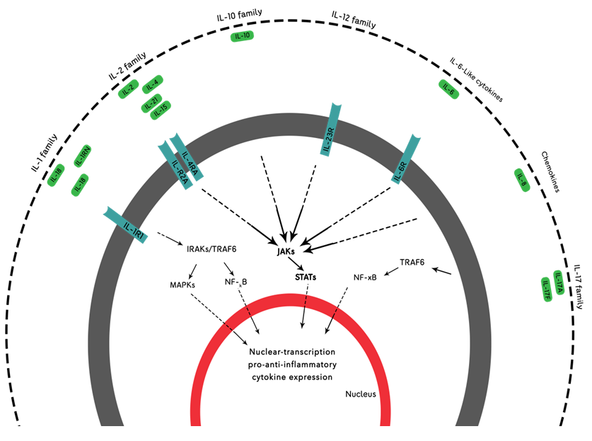
Rheumatoid arthritis is chronic illness caused due to breakdown of immune system which causes severe pain, stiffness and inflammation of joints and it exist in 1% of population. B cells, T cells, macrophages and immune cells gather in swollen joints of patient ; . When we talk about the factors which have significant part in Rheumatoid arthritis disease, the 1st one is hereditary factors which play about 60% role while the other factors like environment, smoking, abnormalities of immune system and obesity also play key role .There are number of interleukins which play a vital role in the inflammation of joints and are present in higher amounts within synovial fluid of patient like IL-1, IL-6, IL-8, IL-17, IL-20, lL-24, IL-19 and IL-35 have anti-inflammatory role in joints ; ; . Tumor necrosis factor also have diverse effects in RA patients that it can induce diabetes mellitus which can leads toward many cardiovascular diseases (IM, 2017). Different cytokines and their receptor, structure, cell source along with their associated disease and their main functions have been shown in Table 1. Many cytokines specially IL-6 and IL-21 induce B cells that produce more than 2% memory cells (CD19, CD21, CD27) which causes increase in the plasma cell production . The inflammatory cytokines such as IL-17 and IL-32 are associated with each other in a way that their productions and functions are interconnected . Different interleukin families that are possibly associated with rheumatoid arthritis disease have been shown in Figure 1.
IL-1
IL-1 and TNF, found high in concentrations within synovial fluid of joints, play critical role in systematic and local inflammation of RA. IL-1 and TNF stimulate conchondrocyts, osteoclast and synoviocyts and these cause destruction in joints ; . The basic part of IL-1 has been seen in initiation of many inflammatory and immune responses and it effects the cell differentiation and proliferation in RA . IL-1B and TNF are shown in initial stages of RA . Meta analysis reveals IL-1B 3954 polymorphism is linked with high possibility of RA . IL-1 have a huge family of cytokines including anti-inflammatory and pro-inflammatory named according to their discoveries from IL-1F1 to IL-1F11 and all of these bind to same receptors comprised of extra cellular immunological domains and intracellular toll/IL-1 domains . IL-33 ST2 is also part of IL-1 family, found in high amount at inflammatory sites released by many immune cells like basophiles NK-cells, eosinophils, mast cells and dendritic cells and directly relates with disease activation and bone erosion . NLRP-3 inflammasome pyrin domain contain 3 induce caspase-1 which produce the IL-1B playing role in inflammation T2D, gout, arthrosecrolosis, mechanical damage and minor trauma in obese RA patients. Number of studies has shown that genetic knock out of NLRP-3 from mice increase insulin sensitivity ; . Furthermore, the inflammatory cytokines especially IL-1B highly correlates with development of cardiovascular diseases and type 2 diabetes in RA patients via the increment of insulin resistance, oxidative stress, dysfunction of endothelial, change in lipid levels and vessel abnormalities so that these patients show more mortality and morbidity rates than the normal RA patients . Studies are still going to treat the RA and T2D by blocking IL-1B, many effective results have gained by using canakinumab (monoclonal antibody) against the IL-1 and anakinara .
IL-6
IL-6 is a pro inflammatory, found in higher amounts in the synovial fluid in various diseases including RA which is formed by several cells together with B cells, T cells, monocytes, endothelial cells and fibroblast cells ; . IL-6 is produced because of infections and injuries and it activates the immune system for protection of body but it’s over production creates many conditions that turn the defensive system against body like in case of RA . In 2008, Tocilizumab (TCB) a humanize monoclonal antibody containing two receptors MIL-6R and SIL-6R, was developed to treat RA with mono therapy as TCB is an IL-6 inhibitor. Its safety and efficacy is more similar to TNF inhibitors. In 2017, it was proven effective against many diseases, moreover, IL-6 deficiency completely eradicates the RA. The normal intimal lining becomes thick due to the production of bone marrow driven macrophages like synoviocytes and fibroblast like B synoviocytes (FLS) which are produced by TNF and IL-6. Many arthritis symptoms are produced because of the production of many bioactive substances like MMP, VEGF, RANKL and INF by Activated FLS which in turns produce fatigue, anemia, pain, bone erosion, cartilage destruction and swallowing ; ; . In mice model, the knocking out IL-6 causes reduction in bone and cartilage damage even though TNF and IL-1B is consistently releases . TH-17 can stimulate many pro inflammatory chemokines, cytokines and macrophages and results in joints damage and inflammation which can be reduced or blocked by the blockage of IL-6 and IL-21 thus helps in the improvement of disease. CD4+ cells contribute to 86% of TH-17 development when both IL-6 and IL-21 are involved while if IL-6 is absent then only 46% T cells reaches to TH-17 and if both are blocked then only 17% TH-17 produced ; . Furthermore, the mice with low level IL-6 generate less amounts of TH-17 and suppression of Treg can also induce higher amounts of TH-17. IL-6, IL-1 along with TNF are produced by CD4+ which produce autoreactive B cells which activate the macrophages and plasma cells to produce these cytokines that further leads to stimulates many chemokines, NK, mast cells, B cells, dendritic cells along with plasma cell and all these contribute in the progression of RA .
IL-17
Interleukin-17 is pro inflammatory which has key role in rheumatoid arthritis and also found in high concentration in synovial fluid of patients. It induces chemokines, cytokines and number of other inflammatory cells which cause damage to joints. IL-17, TNF- α and IFN are found in early stages of inflammation of RA and these induce the stimulation of macrophages . IL-7R also have major role in the pathology of disease. It can induce immune cells due the presence of receptors protein involved in it. Five SNPS of IL-7R have crucial role in RA but the two rs969129 and 6451231 play the major role . IL-17 and IL-32 are linked to each other such that they affect each other’s production and both of these have role in osteoclastogenesis in RA ; . Suppression of IL-17 and TH-17 can reduce the inflammation in RA when we inject the recombinant IL-37 or adenovirus mediated IL-37. IL-37 cannot perform direct inhibition of pro inflammatory cytokine IL-36 however, it binds with IL-1R8 which interrelates to IRAK and TRAF6 then inhibits IL-1, IL-36, IL-18, IL-33 and TLR . The obese patient with RA shows high level of IL-17A and IL-17F in TH17 cells which amplify the fat-oriented inflammation and it has been seen that adipose tissues in obese RA patients secrete many pro inflammatory cytokines along with cells which cause injure to joints and worsen the disease . Bone erosion cause the bone damage and softening through the induction of osteoclast and blockage of these osteoblast will helps in the bone formation because of the emission of pro inflammatory cytokines like IL-1, TNF Alpha and IL17 . TH-17 produces IL-17 causing inflammation and recruitment of leucocytes at the place of inflammation . In 2003, a study was shown on RA models of animals, CD4+ cells which are distinguished with activation of IL-17 and proliferation due to IL-23, have crucial role in collagen induce arthritis activation and joint damage known as TH17. TH17 plays vital part in RA pathogenesis by producing many chemokines and cytokines like IL-6, IL-8, GM-CFS and CC chemokine ligand (CCL)-20 .
IL-21
IL-21 is pro inflammatory cytokine family member of cytokine 1 and found in high level in serum and SF. It increases the production of B cells which produce ACPA and IL-6 in RA patients. IL-21 boosts the production of plasma-blasts whereas the B cells activate the many other pro inflammatory cytokines, auto antibodies and antigen presenting cells which contribute in expansion of disease. Many studies on murine models showed the crucial role of IL-21, for example, trails with K/NxB IL-21R in mouse and IL-21 Rfc fusion protein cause delayed in the initiation and reduced the rigorousness of the arthritis ; . IL-21 activates the production of T follicular helper cells via Bcl-6 increment, CD4+, CD8+ and NK, dendrite cells, fibroblast cells and plamoblast cells through the combine signals of receptor gamma chain and IL-21R which contributes in the severity of disease. Tfh cell promotes the propagation of B cells by going into germinal center, along with antibody and IgG production which correlates to the secondary immunization in mice R.KO. Studies have shown that the reduction or absence of IL-21 can reduce the transmission of GC into B cells which are the main resource of RF and anti-ccp antibodies which contributes in the autoimmune syndromes like RA . TH17 cell production also relates with the IL-6 and IL-21. Experiments have shown that if we block both IL-6 and IL-21 in mouse having CIA, AIA and RA then they result in reduction of TH17 differentiation which relates with the pathogenesis of RA. Furthermore these both cytokines are also involved in the making of IL-23 that also promote the separation of TH17 and which further promotes many other cytokines like IL-17, IL-21, IL-22 involved in progression of arthritis .
Table 1: Different cytokines and their receptor, structure, cell source along with their associated diseases and their main functions (Akdis et al., 2016).
| Cytokines | Receptors | Structure | Cell source | Associated disease | Main functions |
| IL-1α, IL-1β | IL-1RI, IL-1RII | Heterodimer | Monocytes, macrophages, neutrophils, synovial lining, fibroblast, lymphocytes | RA, Psoriasis, IBD and many other inflammatory diseases | Hematopoiesis, TH17 cells differentiation, pro inflammatory protein induction |
| IL-6 | IL-6Ro (Sil-6R) GP130 | homodimer | Monocytes, endothelial cells, macrophages, fibroblast | SLE, RA, Chronic inflammatio, autoimmune diseases, castle man disease | B and T cells differentiation IgA, IgG, IgM hematopoiesis |
| IL-17A, IL-17F | IL-17RA, IL17RC | Heterodimer, homodimer | CD4+, Th17 cells, NK Cells, T cells, NKT cells, neutrophils | IBD, RA, allergic asthma, MS, psoriasis | Pro inflammatory chemokine and cytokine induction, neutrophils recruitment |
| IL-21 | IL-21R | Monomer, 4 helix bundle | NKT cells, T cells, TH17 specially | RA, cancer, SLA | Proliferation regulation, balance of antibody isotype differentiation |
| IL-27 | Gp130, wsx-1 | Heterodimer | Activated DCs, epithelial cells, macrophages | Immune pathology | TH17 inhibition via STAT1, Tbet induction, promoting TH1 cells |
| IL-32 | Unclear | Unclear | NK, monocytes, epithelial cells, T cells, macrophages, | Inflammatory bowel disease, RA, Cancer, crohn disease | Induction of IL-8, TNF-α, IL-6 and epithelial cells apoptosis |
| IL-35 | Unclear | Heterodimer | Treg cells | Collagen induce arthritis, IBD | TH17 cells inhibitor, Treg proliferation and inflammatory response suppression |
| IL-37 | IL-18RA | Unclear | DC cells and intra cellular mechanisms manner | RA | DC inhibitor and inflammatory cytokine suppression |
IL-27
IL-27, belonging from IL-12 has anti-inflammatory activity and is found in high amount in the synovial fluid derived from CD14+ MNCs. It has been noticed that IL-27 can reduce the formation of many pro inflammatory cytokines including IL-6 so it is supposed that it has anti-inflammatory effect on RA. IL-27 can reduce production of TH2 and TH17 through releasing INF and TH1 so that it can inhibit the GATA-3 and generates the T-bet cells . Higher levels of IL-27 can induce proteoglycan arthritis in murine models and adjuvant induced arthritis in rat along with autoimmune encephalomyelitis in rats. FLS are produced by the IL-27 which further induce many other inflammatory chemokines including CCL2, CXC9, CXC1 and cytokines IL-6, IL-12, IL-17, IL-21, TNF alpha and INF gamma via the stimulation of many intracellular pathways like c-Jun amino-terminal kinase (JNK) and p38 mitogen activated protein kinase (MAPK) along with extracellular signal-regulated protein kinase (ERK) which can also induce IL-6, IL1B and TNF along with MMP which cause pannus formation and cause damage to tissues and cartilage. VCAM-1 and ICAM-1 are the hallmark of the RA, also expressed on FLS can activate infiltration of leukocytes joints . Many studies show the contrary role of IL-27 as it activates T cells and INF gamma shows its inflammatory characteristic in murine models which have reduce Treg and some studies also suggest that the injection of IL-27 reduce the rigorousness of infection and osteoclastogenesis in murine model of CIA via the down regulation TH17 and up regulation of Treg . Many studies in murine model have revealed that IL-17 proliferated by TH17 is involved in pathogenesis of RA using the joint destruction and inflammation can be reduced by the introduction IL-27 through STAT3/ROR gammaT . IL-27 can increase the level of ESR, CRP anti-CCP by the induction of many inflammatory cells like mast cells, NK, mononuclear cells, TH-1 and T cells which further involve in formation of cytokines like IL-1, IL-12, IL-18, IFN gamma and TNF alpha so that they are not accurate markers for the RA disease. IL-27 can block the production of TH17 at early stages and promotes the generation of Treg and, IL-10 and IL-17 in CD4+ T cells. Its role in pathogenesis of disease is still unclear but it blocks the inflammatory mediators, tissue damage, cell survival, apoptosis and angiogenesis and reduces inflammation. IL-27 can be used as a biomarker for the RA disease as it has high levels and correlates with DAS28 .
IL-32
The newly discovered interleukin, its gene is found on chromosomal location 16p13.3 and there are nine isoforms with different properties and activities referred as mRNA known as NK transcript 4 (NK4) has similarity with protein produced by IL-2 NK cells, and this is produced by both immuned and non immuned cells and have no similarity to other identified cytokines. IL-32 gains attention due to its biological properties. IL-32 has strong association with TNF alpha and can stimulate many pro inflammatory cytokines (IL-6, TNF alpha, IL12, IL-1β, TH1 and TH17) by NK-KB and p38MAPK ; . IL-32 is formed in many different cells including NK cells, T cells, epithelial cells and monocytes. Injections of IL-32 cause inflammation, damage of joints and infiltration in mice models revealing its pro inflammatory properties. IL-17 and IL-32 link together and affect each other production. In addition to this, IL-32 production is also linked with the TNF alpha and studies have shown that treatment with anti-TNF alpha cause reduction in IL-32. It also produces osteoclaste which damage the joint and play crucial role in osteoclastogenesis .
IL-35
IL-35, member of class IL-12 have two subunits EB13 and p35, found in elevated levels in rheumatoid arthritis . The function of IL-35 is contentious, several studies show its anti-inflammatory roles and some show its inflammatory roles in inflammatory disease including RA. IL-35 encourages production of Treg cells and inhibits the TH17 which perform key part in segregation of osteoclasts and it depends on the RANKL signaling pathway that is correlated with IL-17. IL-35 suppresses the IL-17 and RANKL signaling pathway that reduce the arthritis in mice models and FSL culturing cells, it also enhances the expression of OPS which act as blocker of RANKL . It reduces the bone and cartilage damage by decreasing osteoclast expression. It can also inhibit the growth and promotes apoptosis of FLS. IL-35 can induce many pro inflammatory cytokines (IL-1B, IL-6, MCPs) when TNF alpha stimulates the IL-35 and EB13 in FSL and MCs (Alunno et al., 2017; ; . As IL-35 is member of IL-12 that perform main part in pathology of RA found in elevated levels in serum relates with systematic scrolesis and in circulation, relates to different types of cancer. Surprisingly, IL-35 levels are found in decreased amount in provocative bowel infection and systematic lupus erythematosus in comparison with control group . The study shows the possible links between the disease activity and level of Treg and IL-35 that is lower than the healthy control which suggest that it is an inflammatory cytokine in RA. The levels of these both also correlates negatively with the DAS28 and ESR which reflects that it is a possible indicator of the disease activity but it does not correlates with the CRP, CCP and RF . Many studies show that the chance of arthritis development could be reduced by introduction of Treg derived from induce pluripotent stem cells .
IL-37
IL-37 is family member of IL-1 also known as IL1F7, acts as basic inhibitor of innate immunity found at human chromosome 2q13. Pro inflammatory cytokines could induce IL37 which suppress the pro inflammatory cytokines like IL-6, TNF, INF-ϒ, IL-18 and IL-1B. IL-37 down regulates or inhibits the expression of IL-17 and Th17 which shows that it have great association to pathology of disease ; ; . Several studies have been conducted on IL-37 to check either it can help to eradicate the disease due to its anti-inflammatory property. Several studies show that IL-37 expression can be lowered by the use of anti-TNF and DMARD treatments because its expression is highly correlated to several anti and pro inflammatory cytokines . T-cell immunoglobulin and mucin domain-containing-3 (Tim-3) are involved in Th-1 mediated immunity related to formation of TNF alpha that has role in pathogenesis of RA and links with the IL-37 production. Their relation is still unclear but IL-37 production reduces the pathogenesis and correlates with the cytokines IL-6 and TNF production but it cannot completely eradicate the disease . Tim-3 expresses on various T cells surfaces that contributes in the pathogenesis of RA like CD3+CD4+ T cells and CD3+CD8+ T cells misbalance can induces RA but CD4+ plays most essential part in activation of RA and CD8+ have no role in the pathogenesis. Studies on animal models show that the reduction in the CD8+ can initiate and transfer of arthritis in sever combine immune deficiency (SCID) . Five different variants of IL-37 (IL-1F7a-e) have been found and the largest isoform is IL-1F7b that has similar homology sequence with IL-18 and is not recognized as anagonist to IL-18 because it can easily binds to IL-18Rα (IL-18 receptor α chain) .
Neuroimmune interactions in rheumatoid arthritis
Neuroimmune cross talk is somewhat latest area of studies which examines effect of neuroimmune cross talk on the inflammation processes of rheumatoid arthritis (RA). Immune functions in the body are regulated all the way through vagus nerve and hypothalamic-pituitary-adrenal (HPA) axis. The vagus nerve, through the cholinergic anti-inflammatory pathway, manages cytokine formation and inhibits pro-inflammatory intermediaries such as TNF-α, IL-1β, and IL-6 synthesis. In people suffering from rheumatoid arthritis, this pathway malfunction results in relentless inflammation . The HPA axis is also central to maintaining immune homeostasis because it is responsible for releasing glucocorticoids. Chronic stress or dysregulation of this axis tends to change the secretion rhythm of cytokines and intensifies inflammation together with joint damage .
Responses of the immune systems in RA are also shaped by neurotransmitters norepinephrine and substance P. The presence of substance P in the synovial fluid is associated with increased pain and inflammation of joints because it induces IL-6 and TNF-α production . Norepinephrine is often described as having an immunosuppressive effect. However, in situations of chronic stress, norepinephrine shifts T-cell activity and increases pro-inflammatory cytokine secretion, which boosts inflammation . The underlying mechanisms of several cytokines pertaining to rheumatoid arthritis (RA) also overlap with neural pathways. Specifically for RA, IL-1β and IL-6 are known to activate glial cells in the central nervous system (CNS) which in turn to contributes to neuroinflammation, fatigue, and heightened hyperalgesia, or pain perception . In addition, the actions of TNF-α are not limited to inflammation of the synovium, but also include peripheral depression and cognitive dysfunction, and more inclusively, the complex intertwines of immune dysfunction and neural circuitry . In light of these observations, some novel approaches to treatment are being developed. More anecdotally, vagus nerve stimulus aids in reduction of inflammatory cytokines, and thus, presents a potential non-pharmacological treatment for RA . Stress relief techniques such as cognitive behavioral therapy (CBT), mindfulness, and meditation also stand to lower inflammation through modification of HPA axis along with sympathetic nervous system . Bridging the gap of understanding about the nervous system and immune system functioning in RA could pave the way for dual target treatment options that address joint pathology and systemic symptoms.
Conclusion
The diagnosis and multi-faceted clinical presentation of rheumatoid arthritis (RA) is consequence of multifactorial secrets underlying the intertwining relationship of the immune system and the nervous system. Neuroimmune system disruptions like the cholinergic anti-inflammatory response of vagus nerve and HPA axis involvement are major contributors of relentless inflammation and worsening of the disease. In addition, mediators of inflammation at peripheral level norepinephrine, substance P and proinflammatory cytokines IL-1β, IL-6 and TNF-α, not only fuel synovial inflammation but also modulate central nervous system activity, intensifying pain, fatigue, and cognitive decline.
Implementing the described mechanisms of neuroimmunology provides possibilities for new treatments. Modifying the activity of the vagus nerve using bioelectronic medicine is an example of non-drug treatment as well as restoring balance to the HPA axis. Alongside primary interventions, modification of chronic stress and immune dysregulation can be achieved through cognitive behavioral therapy and mindfulness techniques. Further studies are needed to address both the inflammatory and neurological aspects of RA to formulate effective treatment plans and enhance the patient’s health and well-being.
Declarations
Acknowledgement
Department of Biotechnology, University of Sargodha is highly acknowledged for providing support to conduct this study.
Funding
The study received no external funding.
IRB approval
Not applicable
Ethical statement
Not applicable.
Declaration of generative AI and AI-assisted technologies in the writing process
No generative AI and AI-assisted technologies were used in the writing process.
Conflict of interest
The authors have declared no conflict of interest.
Figures

A schematic diagram showing different receptors and interleukin families that take part in pathogenesis during rheumatoid arthritis. The interleukin receptors or interleukins that show positive relationship between SNPs/genes and phenotype of disease are shown. On the basis of functional properties, receptor chain similarities and sequence homology interleukins are given to each of the family. Moreover, overlap between such families can exist. In rheumatoid arthritis the Polymorphisms in genes that encodes for interleukins and interleukin receptors are involved. The binding of ligand starts phosphorylation of intracellular cascades and are intervened through different kinases including IRAK (interleukin 1 receptor associated kinase), MAPK (mitogen-activated protein kinase), JAK (Janus kinase), TNF (tumor necrosis factor receptor associated factor) and TRAF. These kinases result in transduction of signals by specific transcription factors [Mainly STAT (signal transducers and activators of transcription), NF-κB (nuclear factor kappa light chain enhancer of activated B cells)]. Several anti-inflammatory and pro inflammatory cytokine genes that are involved in Pathogenesis are stimulated by above mentioned transcription factors. (Modified from Magyari et al., 2014).
References
- Abdelal, I.T., Zakaria, M.A., Sharaf, D.M. and Elakad, G.M., 2016. levels of plasma cell-free DNA and its correlation with disease activity in rheumatoid arthritis and systemic lupus erythematous patients. Egypt. Rheumatol., 38: 295-300. https://doi.org/10.1016/j.ejr.2016.06.005[CrossRef]
- Akdis, M., Aab, A., Altunbulakli, C., Azkur, K., Costa, R.A., Crameri, R. and Frei, R., 2016. Interleukins (from IL-1 to IL-38), interferons, transforming growth factor β, and TNF-α: Receptors, functions, and roles in diseases. J. Allergy Clin. Immunol., 138: 984-1010. https://doi.org/10.1016/j.jaci.2016.06.033[CrossRef]
- Alshevskaya, A.A., Lopatnikova, J.A., Shkaruba, N.S., Chumasova, O.A., Sizikov, A.E., Karaulov, A.V. and Sennikov, S.V., 2015. Differnce of IL-1β receptors expression by immunocompetent cells subsets in rheumatoid arthritis. Mediat. Inflamm., 2015. https://doi.org/10.1155/2015/948393[CrossRef]
- Alunno, A., Carubbi, F., Giacomelli, R. and Gerli, R., 2017. Cytokines in the pathogenesis of rheumatoid arthritis: new player and therapeutic target. BMC Rheumatol., 1: 3. https://doi.org/10.1186/s41927-017-0001-8[CrossRef]
- Bai, M., He, Y., Yuan, D., Jin, T. and Wang, L., 2019. IL-7R gene polymorphisms among patients with rheumatoid arthritis: A case control study. Mol. Genet. Genom. Med., 7: e00738. https://doi.org/10.1002/mgg3.738[CrossRef]
- Boutet, M.A., Nerviani, A. and Pitzalis, C., 2019. IL-36, IL-37 and IL-38 cytokines in skin and joint inflammation: A comprehensive review of their therapeutic potential. Int. J. Mol. Sci., 20: 1257. https://doi.org/10.3390/ijms20061257[CrossRef]
- Bremer, L., 2017. Hyaluranan (HA) fragments and initiators or enhancer of inflammation in arthritis patients. Masters thesis. KTH, School of Biotechnology (BIO).
- Burridge, I., Eastham, S., Hill, D., Andrews, R., Szomolay, B., Williams, N. and Jones, S., 2023. Transcriptomic analysis of synovitis identifies inhibition of CXCL1 as a mechanism contributing to resolution of inflammatory arthritis under IL-27 control. https://doi.org/10.1093/rheumatology/kead104.171[CrossRef]
- Jones, G., 2023. P130 Rheumatology. https://doi.org/10.1093/rheumatology/kead104.171.[CrossRef]
- Dam, E.M., Maier, A.C., Hocking, A.M., Carlin, J., Ng, B. and Buckner, J.H., 2018. Increase binding of specificity protein 1 to the IL21R promoter in B cells results in enhance B cell response in rheumatoid arthritis. Front. Immunol., 9: 1978. https://doi.org/10.3389/fimmu.2018.01978[CrossRef]
- Dantzer, R., O’Connor, J.C., Lawson, M.A. and Kelley, K.W., 2022. Inflammation-associated depression: From serotonin to kynurenine. Psychoneuroendocrinology, 137: 105676.
- Felger, J.C., 2022. Role of inflammation in depression and treatment implications. Handb. Clin. Neurol., 198: 177-193.
- Ferraccioli, G., Bracci-Laudiero, L., Alivernini, S., Gremese, E., Tolusso, B. and De Benedetti, F., 2010. Interleukin-1β and interleukin-6 in arthritis animal models: roles in the early phase of transition from acute to chronic inflammation and relevance for human rheumatoid arthritis. Mol. Med., 16: 552-557. https://doi.org/10.2119/molmed.2010.00067[CrossRef]
- Guo, Q., Wang, Y., Xu, D., Nossent, J., Pavlos, N.J. and Xu, J., 2018. Rheumatoid arthritis: Pathological mechanisms and modern pharmacologic therapies. Bone Res., 6: 1-14. https://doi.org/10.1038/s41413-018-0016-9[CrossRef]
- IM, J.H., 2017. Patients with newly diagnosed rheumatoid arthritis are at increased risk of diabetes mellitus an observational cohort study. Acta Reumatol. Port., 4.
- Irwin, M.R. and Slavich, G.M., 2023. Psychosocial stress, inflammation, and depression: Pathways and mechanisms. Trends Cognit. Sci., 27: 12-28.
- Jang, S., Kwon, E. and Lee, J., 2022. Rheumatoid arthritis: Pathogenic roles of diverse immune cells. Int. J. Mol. Sci., 23. https://doi.org/10.3390/ijms23020905[CrossRef]
- Kamiya, Y., Kikuchi, T., Goto, H., Okabe, I., Takayanagi, Y., Suzuki, Y., Sawada, N., Okabe, T., Suzuki, Y., Kondo, S., Hayashi, J. and Mitani, A., 2020. IL-35 and RANKL synergistically induce osteoclastogenesis in RAW264 mouse monocytic cells. Int. J. Mol. Sci., 21. https://doi.org/10.3390/ijms21062069[CrossRef]
- Koopman, F.A., van Maanen, M.A., Vervoordeldonk, M.J. and Tak, P.P., 2021. Balancing the autonomic nervous system to treat chronic inflammation in rheumatoid arthritis. Nat. Rev. Rheumatol., 17: 283-297.
- Kunwar, S., Dahal, K. and Sharma, S., 2016. Anti-IL-17 therapy in treatment of rheumatoid arthritis a systematic literature review and meta anlysis of randomized controlled trials. Rheumat. Int., 36(8): 1065-1075. https://doi.org/10.1007/s00296-016-3480-9[CrossRef]
- Lai, X., Wang, H., Cao, J., Li, Y., Dai, Y., Xiang, Y. and Zhang, L., 2016. Circulating IL-27 is elevated in rheumatoid arthritis patients. Molecules, 21: 1565. https://doi.org/10.3390/molecules21111565[CrossRef]
- Lee, Y.D., Ji, J.D. and Song, G.G., 2009. Association between interleukin 1 polymorphisms and rheumatoid arthritis susceptibility: A meta analysis. J. Rheumatol., 36: 12-15. https://doi.org/10.3899/jrheum.080450[CrossRef]
- Li, Y., Li, D., Wu, S., Jiang, S., Lin, T., Xia, L. and Lu, J., 2016. Interleukin-35 upregulates OPG and inhibits RANKL in mice with collagen-induce arthritis and fibroblast-like synoviocytes. Osteop. Int., 27: 1537-1546. https://doi.org/10.1007/s00198-015-3410-9[CrossRef]
- Liu, R., Wu, Q., Su, D., Che, N., Chen, H., Geng, L. and Sun, L., 2012. A regulatory effect of IL-21 on T follicular helper like cell and B cell in rheumatoid arthritis. Arthrit. Res. Ther., 14: R255. https://doi.org/10.1186/ar4100[CrossRef]
- Liu, Y., Li, Z., Su, H., Zhou, S. and Huang, X., 2023. The role of HPA axis dysregulation in rheumatoid arthritis: From neuroimmune interaction to clinical management. Front. Immunol., 14: 1189456.
- Magyari, L., Varszegi, D., Kovesdi, E., Sarlos, P., Farago, B., Javorrhazy, A. and Meleg, B., 2014. Interleukins and interleukin receptors in rheumatoid arthritis: Research, diagnosis and clinical implications. World J. Orthop., 5: 516. https://doi.org/10.5312/wjo.v5.i4.516[CrossRef]
- Migliorini, P., Italiani, P., Pratesi, F., Puxeddu, I. and Boraschi, D., 2020. The IL-1 family cytokines and receptors in autoimmune diseases. Autoimm. Rev., 102617. https://doi.org/10.1016/j.autrev.2020.102617[CrossRef]
- Miller, A.H. and Raison, C.L., 2023. The role of inflammation in stress-related psychiatric disorders: Mechanisms and therapeutic implications. Ann. Rev. Med., 74: 141-157.
- Moon, S.J., Park, J.S., Heo, Y.J., Kang, C.M., Kim, E.K., Lim, M.A. and Park, S.H., 2013. In vivo action of IL-27: Reciprocal regulation of TH17 and Treg cells in collagen induced arthritis. Exp. Mol. Med., 45: E46-e46. https://doi.org/10.1038/emm.2013.89[CrossRef]
- Moon, Y.M., Yoon, B.Y., Her, Y.M., Oh, H.J., Lee, J.S., Kim, K.W. and Kim, H.Y., 2012. IL-32 and IL-17 interact and have the potential to aggravate osteoclastogenesis in rheumatoid arthritis. Arthrit. Res. Ther., 14: R246. https://doi.org/10.1186/ar4089[CrossRef]
- Narazaki, M. and Kishimoto, T., 2018. The two-faced cytokine IL-6 in host defense and disease. Int. J. Mol. Sci., 19: 3528. https://doi.org/10.3390/ijms19113528[CrossRef]
- Ngcobo, S., Nkambule, B., Nyambuya, T., Mokgalaboni, K., Ntsethe, A., Mxinwa, V., Ziqubu, K., Ntamo, Y., Nyawo, T. and Dludla, P., 2022. Activated monocytes as a therapeutic target to attenuate vascular inflammation and lower cardiovascular disease-risk in patients with type 2 diabetes: A systematic review of preclinical and clinical studies. Biomed. Pharmacother. Biomed. Pharmacother., 146: 112579. https://doi.org/10.1016/j.biopha.2021.112579[CrossRef]
- Ogata, A., Kato, Y., Higa, S. and Yoshizaki, K., 2019. IL-6 inhibitor for the treatment of rheumatoid arthritis: A comprehensive review. Modern Rheumatol., 29: 258-267. https://doi.org/10.1080/14397595.2018.1546357[CrossRef]
- Patel, S., Malhotra, N., Sharma, V. and Tripathi, T., 2021. Substance P and its emerging role in inflammation and immune modulation. J. Immunol. Res., 2021: 6614563.
- Pavlov, V.A. and Tracey, K.J., 2022. The cholinergic anti-inflammatory pathway and neuro-immune interactions. Nat. Rev. Neurosci., 23: 383-396.
- Roeleveld, D.M., Marjinissen, R.J., Walgreen, B., Helsen, M.M., van den Bersselaar, L., van de Loo, F.A. and Koenders, M.I., 2017. Higher efficacy of anti-IL-6/IL-21 combination therapy compared to monotherapy in induction phase of TH17-driven experimental arthritis. PLoS One, 12: e0171757. https://doi.org/10.1371/journal.pone.0171757[CrossRef]
- Roy, T. and Ghosh, S., 2013. Animal models of rheumatoid arthritis: correlation and usefulness with human rheumatoid arthritis. Indo Am. J. Pharm. Res., 3: 6131-6142.
- Ruscitti, P., Cipriani, P., Di Benedetto, P., Liakouli, V., Berardicurti, O., Carubbi, F. and Giacomelli, R., 2015. Monocytes from patients with rheumatoid arthritis and type 2 diabetes mellitus display an increased production of interleukin (IL)-1β via the nucleotide-binding domain and leucin-rich repeat containing family pyrin 3(NLRP3)-inflammasome activation: A possible implication for therapeutic decision in these patients. Clin. Exp. Immunol., 182: 35-44. https://doi.org/10.1111/cei.12667[CrossRef]
- Schinnerling, K., Aguillon, J.C., Catalan, D. and Soto, L., 2017. The role of interleukin-6 signaling and its therapeutic blockage in skewing the T cell balance in rheumatoid arthritis. Clin. Exp. Immunol., 189: 12-20. https://doi.org/10.1111/cei.12966[CrossRef]
- Senolt, L., Sumova, B., Jandova, R., Hulejova, H., Mann, H., Pavelka, K. and Filkova, M., 2015. Interleukin 35 synovial fluid level are associated with disease activity of rheumatoid arthritis. PLoS One, 10. https://doi.org/10.1371/journal.pone.0132674[CrossRef]
- Shahrara, S., Huang, Q., Mandekin, A.M. and Pope, R.M., 2008. TH17 cells in rheumatoid arthritis. Arthrit. Res. Ther., 10: 93. https://doi.org/10.1186/ar2477[CrossRef]
- Shoda, H., Nagafuchi, Y., Tsuchida. Y., Sakurai, K., Sumitomo, S., Fujio, K. and Yamamoto, K., 2017. Increase serum comcentration of IL-1 β, IL-21 and TH17 cells in over weight patients with rheumatoid arthritis. Arthrit. Res. Ther., 19: 111. https://doi.org/10.1186/s13075-017-1308-y[CrossRef]
- Skejoe, C., Hansen, A., Stengaard-Pedersen, K., Junker, P., Hoerslev-Pedersen, K., Hetland, M., Oestergaard, M., Greisen, S., Hvid, M., Deleuran, M. and Deleuran, B., 2022. T-cell immunoglobulin and mucin domain 3 is upregulated in rheumatoid arthritis, but insufficient in controlling inflammation. Am. J. Clin. Exp. Immunol., 11: 34-44.
- Song, L., Wang, Y., Sui, Y., Sun, J., Li, D., Li, D., Li, G. and Shu, Q., 2018. High interleukin-37(IL-37) expression and increased mucin-domain containing -3 (TIM-3) on peripheral T cell in patients with patients. Int. Med. J. Exp. Clin. Res., 24: 5660. https://doi.org/10.12659/MSM.909254[CrossRef]
- Wang, X., Xu, K., Chen, S., Li, Y. and Li, M., 2018. Role if interleukin-37 in inflammatory and autoimmune disease. Iran. J. Immunol., 15: 165-174.
- Wong, C.K., Chen, D.P., Tam, L.S., Li, E.K., Yin, Y.B. and Lam, C.W., 2010. Effects of inflammatory cytokine IL-27 in the activation of fibroblast-like synoviocytes in rheumatoid arthritis. Arthrit. Res. Ther., 12: R129. https://doi.org/10.1186/ar3067[CrossRef]
- Wu, C., Goodall, J.C., Busch, R. and Gaston, J.S.H., 2015. Relationship of CD146 expression to secretion of interleukin (IL)-17, IL-22 and interferon-Ý by CD4+ T cells in patients with inflammatory arthritis. Clin. Exp. Immunol., 179: 378-391. https://doi.org/10.1111/cei.12434[CrossRef]
- Xin, P., Jie, L., Cheng, Q., Bin, D. and Dan, C., 2021. Pathogenesis and function of interleukin-35 in rheumatoid arthritis. Front. Pharmacol., 12. https://doi.org/10.3389/fphar.2021.655114[CrossRef]
- Zhang, X.Y., Zuo, Y., Li, C., Tu, X., Xu, H.J., Gou, J.P. and Mu, R., 2018. IL1F7 gene polymorphism is not associated with rheumatoid arthritis susceptibility in northern Chinese Han population: A case control study. Chinese Med. J., 131: 171. https://doi.org/10.4103/0366-6999.222340[CrossRef]
- Zhang, X., Zhang, X., Zhuang, L., Xu, C., Li, T., Zhang, G. and Liu, Y., 2018. Decreased regulatory T-cell frequency and interleukin-35 level in patients with rheumatoid arthritis. Exp. Therapeut. Med., 16: 5366-5372. https://doi.org/10.3892/etm.2018.6885[CrossRef]
Services
Article usage statistics
Loading…
