Research Article · Volume 10, Issue 7, 20 June 2022 · Open Access
Application of Phages to Control Escherichia coli Infections in Native Noi Chickens
· · · · · ·
Affiliations & Notes ⌄
1Department of Animal Sciences, College of Agriculture, Can Tho University, Can Tho City, Vietnam
2Department of Veterinary Medicine, College of Agriculture, Can Tho University, Can Tho City, Vietnam
3Department of Food Technology, Faculty of Chemical Engineering, Bio and Food Technolgy, Can Tho University of Technology, Can Tho City, Vietnam
4Applied Biology Faculty, Tay Do University, Can Tho City, Vietnam
5School of Agriculture and Aquaculture, Tra Vinh University, Tra Vinh City, Vietnam. *Correspondence | Nguyen Trong Ngu, Department of Animal Sciences, College of Agriculture, Can Tho University, Can Tho City, Vietnam
This study assessed the efficacy of two phages, MHH6 and PR2, against Escherichia coli serotype O6 infected in chickens. A total of 420 broilers were randomly assigned to seven treatment groups. The negative control (NC) birds received no E. coli or phages, whereas the positive control (PC) birds were infected with E. coli only. The NC+MHH6 and NC+PR2 treatments received 109 pfu/ml of phages MHH6 or PR2, respectively; whereas the PC+MHH6, PC+PR2, and PC+MHH6PR2 groups were infected with 107.1 cfu/ml of E. coli strain O6 and treated with 109 pfu/ml of phages of MHH6, PR2, or both. E. coli infection was inoculated in two-day-old chicks, and phages were administered 24 hours later. The mortality rate of chickens in the PC+MHH6PR2 group was significantly lower (16.7%) than in the PC group (58.3%). The frequency of lesions and E. coli densities in the heart, liver, and spleen of chickens treated with MHH6 and PR2 phages were greatly decreased in infected chickens. After 98 days, the body weight of birds from the E. coli-infected groups treated with MHH6 and PR2 phages was lower than individuals in non-E. coli-infected groups (1,313-1,324 g/bird) but higher and significantly different from those in the PC group (1,172 g/bird). The majority of commercially important traits in chickens improved after phage treatment, proving that these phages are capable of controlling O6 E. coli infected in Noi chickens.
Keywords | Bacteriophage, Carcass characteristic, Chicken, E. coli, Growth performance
INTRODUCTION
Increased consumption of livestock products such as meat, milk, and eggs necessitates an increase in production by animal producers to keep up with the demand. As a result, large-scale livestock production systems are continually developing, with high stocking densities facilitating disease transmission and economic losses . Escherichia coli (E. coli) is regularly isolated from poultry intestines and other mucosal surfaces, and it is classed as a harmful bacterium for chicken, capable of producing colibacillosis, a gastrointestinal disease. Antibiotics are frequently used to treat infections caused by bacteria; however, their efficacy is being questioned due to a rising number of antibiotic-resistant bacteria. According to Nhung et al. (2017), the E. coli APEC strains were resistant to ampicillin, amoxicillin, and tetracycline at levels greater than 70% and ciprofloxacin, neomycin, and chloramphenicol at rates between 50% and 70%. Alternative strategies are required in this situation, one of which is using bacteriophages (phages) to fight bacterial infections .
Numerous advantages make phage therapy an appealing option to antibiotics . According to Domingo et al. (2016), phages are selective for certain bacteria, and phage therapy is deemed safe and effective compared to antibiotics. This mechanism of action does not affect the proliferation of gut flora ; . Additionally, because phages are abundant in nature, they can be isolated and selected quickly in opposition to antibiotic synthesis, which requires millions of dollars and years of research to generate an effective antibiotic . Phage supplementation has been proven to increase feed efficiency, body weight gain, pathogen reduction, and egg production in broiler chickens and laying hens . Phage as a feed additive may be an effective way to regulate the gut microbiota of chickens by lowering particular pathogenic microbial populations and increasing beneficial bacteria, resulting in enhanced gut health .
Upadhaya et al. (2021) have established the efficacy of phage therapy at a range of doses, such as 108 pfu/ml and 106 pfu/ml. Their presence aided in decreasing harmful germs in poultry and animals before slaughter. Additionally, phage has been used to extend the life of food packaging materials and as a disinfectant on production lines . Oral administration of a bacteriophage mixture to rats significantly diminished E. coli O157:H7 colonization in the gastrointestinal tract . The phage treatment also significantly reduced the number of E. coli O157:H7 bacteria on the meat surface . However, little study has been undertaken on the efficacy of phage therapy against E. coli infection in indigenous chickens. In a recent study, we isolated two phages, namely MHH6 and PR2, which were shown by the transmission electron microscopy to belong to the Myoviridae family based on their shape and the presence of the tail diameter . These phages could survive at low pH levels and lyse E. coli strains of O1, O78, and O6. Therefore, the current study was undertaken to investigate the effects of MHH6 and PR2 phages on controlling E. coli in native Noi chickens, as measured by their growth performance, carcass characteristics, and the bacteria population in the internal organs.
MATERIALS AND METHODS
Ethical statement
All chickens were handled and cared for in line with Vietnam’s Animal Husbandry Law, No. 32/2018/QH14 dated on December 22, 2018.
Bacteria and phages
In the present study, the E. coli strain of serotype O6 was purchased from ATCC (American Type Culture Collection) (ATCC® 25922TM), and two phages, MHH6 and PR2, were taken from our previous work .
Chickens and experimental procedure
The study was conducted on the experimental farm of Can Tho University located at Campus IV, Phung Hiep district, Hau Giang province. Noi chickens were purchased from Soc Trang breeding company and kept in-house with four cages per treatment. Each cage was outfitted with two drinking troughs and two feeding troughs. Water for drinking was constantly accessible. Commercial feeds were fed ad libitum for two feeding periods from 1 to 28 days (16% crude protein, 4% crude fiber, metabolizable energy 2,800 Kcal/kg) and 29 to 98 days (14% crude protein, 5% crude fiber, metabolizable energy 2,800 Kcal/kg). Following the breeding company’s directions, the vaccination schedule for chickens was thoroughly implemented during the rearing process.
The 98-day experiment included a total of 420 one-day-old Noi broiler chickens. The chicks were randomly assigned to one of seven experimental groups, each with four replicates and 15 broilers per replicate. The treatment groups were: Negative Control (NC) - treatment without E. coli challenge; NC+Bacteriophage 1 (NC+MHH6); NC+Bacteriophage 2 (NC+PR2); Positive Control (PC) - treatment with E. coli challenge; PC+Bacteriophage 1 (PC+MHH6); PC+Bacteriophage 2 (PC+PR2); PC+Bacteriophage 1 and Bacteriophage 2 (PC+MHH6PR2). Chicks were selected for oral infection with 1 ml of E. coli on day two at a dose of 107.1 cfu/ml (based on lethal dose, LD50 results), and phages were administered orally at 109 pfu/ml starting 24 h after infection, followed for three consecutive days and repeated weekly until day 63.
Sampling and measurements
The chicken mortality rate was monitored during the trial. At 7, 21, 35, and 49 days after infection, one chick from each cage was randomly selected and euthanized by exsanguination to determine the density of E. coli and record the weights of internal organs such as heart, liver, and spleen .
Chicken body weight (BW) was determined weekly, and feed intake (FI) was recorded daily for each cage. These data were then utilized to compute body weight gain (BWG) and feed conversion ratio (FCR). At the end of the experiment, 56 chickens (8 per treatment, equal sex) were slaughtered to determine carcass characteristics and organ weights. Breast, thigh, drumstick muscle, and wings were collected according to Faria et al. (2010). In addition, the liver, spleen, heart, and gizzard were removed and weighed, and data were expressed as percentages of body weight.
Statistical analysis
The data were analyzed using the General Linear Model procedure of the Minitab version 16 software (State College, PA, USA) . Tukey’s comparison test with P ≤ 0.05 was performed to detect the mean difference between treatments.
RESULTS and Discussion
The mortality rate of chickens
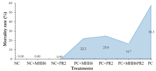
Figure 1 shows the total mortality rate of the experimental chickens. In treatments without E. coli infection (NC) or only with phage treatment (NC+MHH6 and NC+PR2), no dead chickens were found. The highest mortality rate was found in chickens infected solely with E. coli (PC), at 58.3%, followed by PC+PR2 (25.0%) and PC+MHH6 (22.2%) treatments.
The presence of E. coli in the internal organs of Noi chickens
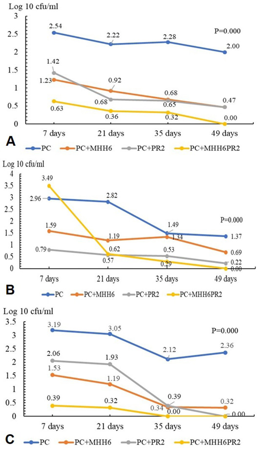
During 49 days of infection, the lytic ability of phages and E. coli invasion were assessed by counting E. coli density in the heart, liver, and spleen of chickens (Figure 2A-C). During the experiment, E. coli were not found in birds’ hearts, liver, and spleen under NC, NC+MHH6, and NC+PR2 treatments. The number of E. coli declined after the 7th day of phage application, and it was eliminated after 49 days in the therapy with both phages included (Figure 2B). Moreover, E. coli in the spleen was deficient and could not be detected in the PC+MHH6PR2 treatment at 35 days or the PR2 phage treatment at 49 days post-infection (Figure 2C). Moreover, the E. coli density in organs of chickens under MHH6 and PR2 treatments was relatively low (0.00-0.69 log10 cfu/ml) compared to the PC group with a higher E. coli population (1.37-2.36 log10 cfu/ml).
Growth performance of chickens
The results in Table 1 indicate that BWG was higher in chickens treated with phages in the first 35 days (258 g/bird) and the whole period (1-98 days) (1,143 g/bird) compared with those in the PC treatment. There was no significant difference in bird growth between the E. coli-free treatments (NC and NC+MHH6; NC+PR2); however, differences were found when treatments with E. coli inoculation were accounted. The overall benefit of improved BWG was also demonstrated in the treatment with both phages included (1,237 g/bird). In addition, higher feed consumption in the non-treated chickens, combined with lower BWG, were responsible for a higher FCR (4.09) in the PC treatment than in the others (3.32-3.71).
Carcass traits of chickens
The phage therapy had a considerable impact on the carcass and organ weight of infected chickens (Table 2). Chickens in the only-phage-treated group, as well as those in a co-inoculation group with E. coli and bacteriophages, had a higher carcass weight and carcass percentage than chickens in the only E. coli-infected group (P<0.05). Additionally, Table 2 demonstrates that the rate of organ weight treated with PC was generally more remarkable than that treated with bacteriophage, particularly the spleen (P<0.01).
Table 1: Effect of bacteriophage on on body weight gain (BWG, g), feed intake (FI, g) and feed converstion ratio (FCR, feed/gain).
Parameters
Treatments
SEM
P
NC
NC+ MHH6
NC+ PR2
PC+ MHH6
PC+ PR2
PC+ MHH6PR2
PC
Feed intake (g)
1-35 d
834abc
816abcd
781cd
798bcd
768d
851ab
871a
14.0
0.000
36-70 d
1,795bc
1,804b
1,776bc
1,738bc
1,679c
1,970a
2,003a
26.4
0.000
71-98 d
1,682ab
1,707ab
1,700ab
1,692ab
1,601b
1,773a
1,805a
29.8
0.003
1-98 d
4,312b
4,328b
4,259b
4,228b
4,049c
4,595a
4,680a
38.6
0.000
Body weight gain (g)
1-35 d
333a
328a
322a
300c
270bc
302b
258d
8.96
0.000
36-70 d
694
698
689
645
687
663
536
40.2
0.105
71-98 d
396
438
439
353
387
396
478
27.2
0.068
1-98 d
1,285a
1,296a
1,284a
1,194c
1,218bc
1,237b
1,143d
7.24
0.000
FCR (feed/ gain)
1-35 d
2.50bc
2.49bc
2.43c
2.66bc
2.84b
2.82b
3.37a
0.08
0.000
36-70 d
2.71b
2.59b
2.58b
2.70b
2.45b
2.97b
3.76a
0.15
0.000
71-98 d
4.27
4.02
3.89
4.92
4.17
4.52
3.80
0.31
0.212
1-98 d
3.35d
3.34d
3.32d
3.54c
3.32d
3.71b
4.09a
0.04
0.000
NC: negative control, without E. coli, without bacteriophage; PC: positive control, E. coli challenged, without bacteriophages; NC+MHH6 and NC+PR2: negative control plus MHH6 or PR2 bacteriophage, respectively; PC+MHH6, PC+PR2: positive control plus MHH6 or PR2 bacteriophage, respectively; PC+MHH6PR2: positive control plus both MHH6 and PR2 bacteriophages. a,b,c,d Within a row, values with different superscripts differ statistically at P<0.05.
Table 2: The effect of bacteriophage supplementation on carcass characterictis and organ weight in broilers.
Parameters
Treatments
SEM
P
NC
NC+ MHH6
NC+ PR2
PC+ MHH6
PC+ PR2
PC+ MHH6PR2
PC
Body weight (BW), g
1,314a
1,324a
1,313a
1,222c
1,247bc
1,266b
1,172d
7.29
0.000
Carcass weight (CW), g
896b
944a
934ab
845c
818c
900ab
654d
9.72
0.000
Carcass, % BW
68.2ab
71.3a
71.1a
69.1ab
65.6b
71.1a
55.8c
0.96
0.000
Breast, % CW
15.7ab
16.3ab
17.2a
15.9ab
14.9b
15.8ab
15.1b
0.38
0.009
Thigh + drumstick, % CW
24.5a
24.7a
24.5a
23.4ab
20.8c
21.6bc
20.2c
0.53
0.000
Wing, % CW
14.0b
14.9ab
15.6a
15.4ab
15.5a
15.1ab
15.3ab
0.31
0.031
Organ weight, % BW
Liver
2.08b
2.18ab
2.13ab
2.37a
1.90b
1.95b
2.12ab
0.06
0.001
Spleen
0.19b
0.19b
0.23ab
0.19b
0.19b
0.21ab
0.27a
0.01
0.006
Gizzard
3.13a
2.39cd
2.67bcd
2.76abc
2.23d
2.98ab
2.77abc
0.010
0.000
Heart
0.55a
0.42b
0.53ab
0.51ab
0.42b
0.52ab
0.52ab
0.03
0.005
NC: negative control, without E. coli, without bacteriophage; PC: positive control, E. coli challenged, without bacteriophages; NC+MHH6 and NC+PR2: negative control plus MHH6 or PR2 bacteriophage, respectively; PC+MHH6, PC+PR2: positive control plus MHH6 or PR2 bacteriophage, respectively; PC+MHH6PR2: positive control plus both MHH6 and PR2 bacteriophages. a,b,c,d Within a row, values with different superscripts differ statistically at P<0.05.
According to Loc-Carrillo and Abedon (2011), a phage is an antibacterial, bactericidal agent that can increase in number during treatment and tends to disrupt the normal flora only to a minimum extent. The ability of phages to host diverse bacterial species reflects their specialization, and the host spectrum of a bacteriophage is defined as the number of bacteria employed by the phage. Therefore, when using phages with a broad host spectrum, it is possible to support the treatment of many strains of bacteria simultaneously. In the in vitro study , MHH6 and PR2 phages demonstrated the wide host range and the ability to lyse O1, O78, and O6 E. coli strains; however, more testing in vivo was required. The data in this study showed that most commercially important traits in chickens improved after phage treatment (Tables 1 and 2). These phages could control O6 E. coli infected in native Noi chickens. At the same time, the mortality rate of the combined treatment of E. coli infected-birds and phage treated-birds was significantly reduced (Figure 1). These findings were consistent with some previous studies, where controlled E. coli bacteria in the intestinal tract of chickens significantly improved carcass quality . Lau et al. (2010) also demonstrated that when phages were used, the mortality rate of chicken was dramatically decreased (P<0.05). At the end of the experiment, the total mortality rate of birds inoculated with 108 cfu of E. coli was 83.3%. When using a phage concentration of 1.5 x 10⁹ pfu/ml, an average decrease of 25% in chicken mortality was noted . Additionally, Naghizadeh et al. (2019) reported that E. coli caused about 46.6% mortality in 15 days old chicks without phage treatment. Only 13.3% of birds died after being injected with 108 cfu E. coli and 1010 pfu of the comparable therapy phage. It can be inferred from these findings that phage therapy effectively inhibits the growth of E. coli and can be utilized to treat E. coli infection in broilers.
Regarding the frequency of lesions and E. coli densities in internal organs of chickens using phages to treat E. coli, Lau et al. (2010) reported that E. coli colonization was reduced in the liver, heart, and spleen and cleared from the blood samples during the experimental period. Utilizing a phage concentration of 1.5 x 10⁹ pfu/ml resulted in a 41.7% reduction in infection incidence compared with the untreated group . Additionally, Lim et al. (2011) showed that after three weeks of follow-up, the rate of bacterial re-isolation in the liver and spleen of hens with just bacterial infections was 40-60%, compared to only 20-40% in the bacteriophage-treated group. It can be explained that phages can move through the mucosal surface and even the blood-brain barrier and effectively protect the host . These findings were consistent with the current study, according to which phage treatment could prevent and reduce the severity of infected chickens.
Colibacillosis is often lethal in chickens, particularly broilers and turkeys. E. coli enters the bloodstream from the site of infection, where it spreads to multiple internal organs, causing sepsis and bird death . Naghizadeh et al. (2019) also discovered a high presence of E. coli in the hearts, livers, and spleens of birds only infected with E. coli (3/8 birds) 10 days post-challenge. However, when using phage for treating, E. coli was reduced in the heart, liver, and spleen after ten days of the challenge. It can be stated that phage administration effectively inhibited E. coli growth in the internal organs of chickens in the present study. According to Bicalho et al. (2010), phages 1230-10 had the highest antibacterial activity and entirely stopped the growth of 71.7% of all isolates. Additionally, the combined action of phage preparations led to a broad spectrum of activity, completely inhibiting 80% of E. coli strains and significantly suppressing the growth of 90% of E. coli isolates. According to Dąbrowska (2019), phages are frequently found in the liver and spleen. The most effective phages are commonly found in these internal organs, even at concentrations more significant than those seen in the blood. Following systemic delivery, the phage can reach the spleen and liver in minutes and get relatively high titers in 1 to 3 hours . Typically, the spleen is the organ where live phages may be identified for the greatest period, even days after phage treatment .
In contrast with the present study (Table 2), Kim et al. (2013) demonstrated that FI and FCR were not affected by phage treatment (0.05%, 0.1%, and 0.2%; 109 pfu/g) to broiler diets. The inclusion of phages at 0.05 and 0.1% in the feed did not affect the FI and FCR of broilers . However, these authors showed a significant increase in BWG with an increase in bacteriophage supplement levels during the starter and slaughter phases. In contrast, Huff et al. (2004) suggested that BWG was not affected by the addition of either DAF6 and SPR02 phages via intramuscular injection (3.7 × 10⁹ and 9.3 × 10⁹ pfu/ml, respectively) in broiler chickens without E. coli infection. Moreover, Lau et al. (2010) discovered that chickens infected with E. coli but not treated with phage had the smallest weights at 14 and 21 days of age. The chicken weight was not significantly different between the E. coli-infected but bacteriophage-treated and the negative control groups.
In the context of the genomes, one may worry about the safety of using phages in chickens or other animal species. This issue is not covered in the present study; however, according to Summers (2001), the majority of phages identified to date are lytic and only a small proportion are capable of integrating their DNA into host chromosomes. Moreover, sequencing can support to eliminate the usage of phages whose genomes encode known harmful products, such as toxins, transposases, or repressor proteins . Previously, Krylov et al. (1993) also pointed out that large-scale manufacturing of phages results in a small proportion of mutated phages. The mutation process typically makes the phage inactive but not stronger, and the most commonly reported mutation renders phages incapable of infecting bacteria. Approximately 10% of phage particles undergo such mutations during large-scale production, but this process often does not compromise phage safety.
CONCLUSIONS and Recommendations
The application of phage therapy in native Noi broilers considerably reduced mortality, as well as the severity of gross lesions in chicken infected with E. coli strain of serotype O6, and contributed to improving growth performance and carcass characteristics of infected birds. This study confirmed the beneficial effects of bacteriophages in controlling E. coli infection in native broilers, which have a slower growth rate but superior meat quality.
ACKNOWLEDGMENTS
This study is funded in part by the Can Tho University Improvement Project VN14-P6, supported by a Japanese ODA loan.
Novelty Statement
The isolated MHH6 and PR2 phages displayed a broad host range and the ability to lyse O1, O78, and O6 E. coli strains yet further in vivo testing still was required. The study revealed that phage treatment in native Noi broilers dramatically reduced mortality and the severity of gross lesions in chickens infected with the serotype O6 E. coli strain, and led to enhanced growth performance in infected birds.
AUTHOR’S CONTRIBUTION
LHA and NTN contributed to the study’s design. The data collection and analysis were carried out by LHA, TTHM, LMT and LTTL. The first draft was prepared by LHA, HTL, and NHX, and the manuscript was corrected by NTN. The final manuscript has been read and approved by all authors.
Conflict of interest
The authors have declared no conflict of interest.
Figures
Figure 1:.
The efficacy of a bacteriophage on mortality rate of chicken E. coli challenged. NC: negative control, without E. coli, without phage; PC: positive control, E. coli challenged, without phages; NC+MHH6 and NC+PR2: negative control plus MHH6 or PR2 phage, respectively; PC+MHH6, PC+PR2: positive control plus MHH6 or PR2 phage, respectively; PC+MHH6PR2: positive control plus both MHH6 and PR2 phages.
Figure 2:.
Number of E. coli bacteria in heart, liver, and spleen organs over 49 days of infection. (a) Heart; (b) Liver; (c) spleen; PC: positive control, E. coli challenged, without phages; PC+MHH6, PC+PR2: positive control plus MHH6 or PR2 phage, respectively; PC+MHH6PR2: positive control plus both MHH6 and PR2 phages.
References
Antao EM, Glodde S, LiG, Sharifi R, Homeier T, Laturnus C, Diehl I, Bethe A, Philipp HC, Preisinger R, Wieler LH, Ewers C (2008). The chicken as a natural model for extraintestinal infections caused by avian pathogenic Escherichia coli (APEC). Microb. Pathog., 45(5-6): 361-369. https://doi.org/10.1016/j.micpath.2008.08.005[CrossRef]
Bicalho RC, Santos TMA, Gilbert RO, Caixeta LS, Teixeira LM, Bicalho MLS, Machado VS (2010). Susceptibility of Escherichia coli isolated from uteri of postpartum dairy cows to antibiotic and environmental bacteriophages. Part I: Isolation and lytic activity estimation of bacteriophages. J. Dairy Sci., 93(1): 93-104. https://doi.org/10.3168/jds.2009-2298[CrossRef]
Cieplak T, Soffer N, Sulakvelidze A, Nielsen DS (2018). A bacteriophage cocktail targeting Escherichia coli reduces E. coli in simulated gut conditions, while preserving a non-targeted representative commensal normal microbiota. Gut. Microb., 9(5): 391-399. https://doi.org/10.1080/19490976 https://doi.org/10.3382/ps/pex359.2018.1447291[CrossRef]
Clavijo V, Florez MJV (2018). The gastrointestinal microbiome and its association with the control of pathogens in broiler chicken production: A review. Poult. Sci., 97(3): 1006-1021. https://doi.org/10.3382/ps/pex359[CrossRef]
Dąbrowska K (2019). Phage therapy: What factors shape phage pharmacokinetics and bioavailability? Systematic and critical review. Med. Res. Rev., 39(5): 2000-2025. https://doi.org/10.1002/med.21572[CrossRef]
Dissanayake U, Ukhanova M, Moye ZD, Sulakvelidze A, Mai V (2019). Bacteriophages reduce pathogenic Escherichia coli counts in mice without distorting gut microbiota. Front. Microbiol., 10: 1984. https://doi.org/10.3389/fmicb.2019.01984[CrossRef]
Domingo-Calap P, Georgel P, Bahram S (2016). Back to the future: bacteriophages as promising therapeutic tools. HLA, 87(3): 133-140. https://doi.org/10.1111/tan.12742[CrossRef]
El-Shibiny A, El-Sahhar S, Adel M (2017). Phage applications for improving food safety and infection control in Egypt. J. Appl. Microbiol., 123(2): 556-567. https://doi.org/10.1111/jam.13500[CrossRef]
Faria PB, Bressan MC, de Souza XR, Rossato LV, Botega LMG, da Gama LT (2010). Carcass and parts yield of broilers reared under a semi-extensive system. Braz. J. Poult. Sci., 12(3): 153-159. https://doi.org/10.1590/S1516-635X2010000300003[CrossRef]
Golkar Z, Bagasra O, Pace DG (2014). Bacteriophage therapy: a potential solution for the antibiotic resistance crisis. J. Infect. Dev. Ctries. 8(2):129-136. https://doi.org/10.3855/jidc.3573[CrossRef]
Gomes A, Quinteiro-Filho W, Ribeiro A, Ferraz-de-Paula V, Pinheiro M, Baskeville E, Akamine A, Astolfi-Ferreira C, Ferreira A, Palermo-Neto J (2014). Overcrowding stress decreases macrophage activity and increases Salmonella Enteritidis invasion in broiler chickens. Avian Pathol., 43(1): 82-90. https://doi.org/10.1080/03079457.2013.874006[CrossRef]
Huff WE, Huff GR, Rath NC, Balog JM, Donoghue AM (2004). Therapeutic efficacy of bacteriophage and Baytril (enrofloxacin) individually and in combination to treat colibacillosis in broilers. J. Poult. Sci., 83: 1944-1947. https://doi.org/10.1093/ps/83.12.1944[CrossRef]
Huh H, Wong S, Jean JS, Slavcev R (2019). Bacteriophage interactions with mammalian tissue: Therapeutic applications. Adv. Drug Deliv. Rev., 145: 4-17. https://doi.org/10.1016/j.addr.2019.01.003[CrossRef]
Isroli I, Yudiarti T, Widiastuti E, Wahyuni HI, Sartono TA, Sugiharto S (2018). Effect of bacillus probiotics on internal organs and carcass characteristics of broiler chicks infected with avian pathogenic Escherichia coli. Livest. Res. Rural Dev., 30(11). http://www.lrrd.org/lrrd30/11/sgh_u30183.html
Kim KH, Lee GY, Jang JC, Kim JE, Kim YY (2013). Evaluation of anti-SE bacteriophage as feed additives to prevent Salmonella enteritidis (SE) in broiler. Asian Austral. J. Anim., 26(3): 386–393. https://doi.org/10.5713/ajas.2012.12138[CrossRef]
Krylov V, Pleteneva E, Shaburova O, Bourkaltseva M, Krylov S, Chesnokova E, Polygach O (2014). Common pre-conditions for safe phage therapy of Pseudomonas aeruginosa infections. Adv. Microbiol., 4: 766-773. https://doi.org/10.4236/aim.2014.412084[CrossRef]
Krylov VN, Tolmachova TO, Akhverdian VZ (1993). DNA homology in species of bacteriophages active on Pseudomonas aeruginosa. Arch. Virol., 131: 141-151. https://doi.org/10.1007/BF01379086[CrossRef]
Lan RX, Lee SI, Kim IH (2017). Effects of Enterococcus faecium SLB 120 on growth performance, blood parameters, relative organ weight, breast muscle meat quality, excreta microbiota shedding, and noxious gas emission in broilers. J. Poult. Sci., 96: 3246-3253. https://doi.org/10.3382/ps/pex101[CrossRef]
Lau GL, Sieo CC, Tan WS, Hair-Bejo M, Jalila A, Ho YW (2010). Efficacy of a bacteriophage isolated from chickens as a therapeutic agent for colibacillosis in broiler chickens. J. Poult. Sci., 89(12): 2589-2596. https://doi.org/10.3382/ps.2010-00904[CrossRef]
Lim TH, Lee DH, Lee YN, Park JK, Youn HN, Kim MS, Lee HJ, Yang SY, Cho YW, Lee JB, Park SY, Choi IS, Song CS (2011). Efficacy of bacteriophage therapy on horizontal transmission of Salmonella Gallinarum on commercial layer chickens. Avian Dis., 55(3): 435-438. https://doi.org/10.1637/9599-111210-Reg.1[CrossRef]
Loc-Carrillo C and Abedon, ST (2011). Pros and cons of phage therapy. Bacteriophage, 1(2): 111-114. https://doi.org/10.4161/bact.1.2.14590[CrossRef]
Lone A, Anany H, Hakeem M, Aguis L, Avdjian AC, Bouget M (2016). Development of prototypes of bioactive packaging materials based on immobilized bacteriophages for control of growth of bacterial pathogens in foods. Int. J. Food Microbiol., 217: 49-58. https://doi.org/10.1016/j.ijfoodmicro.2015.10.011[CrossRef]
Minitab (2010). Minitab reference manual, Release 16.2.1 for Windows. Minitab Inc, USA.
Naghizadeh M, Torshizi MAK, Rahimi S, Dalgaard TS (2019). Synergistic effect of phage therapy using a cocktail rather than a single phage in the control of severe colibacillosis in quails. J. Poult. Sci., 98(2): 653-663. https://doi.org/10.3382/ps/pey414[CrossRef]
Ngu NT, Loc HT, Nhan NTH, Huan PKN, Anh LH, Xuan NH (2020). Isolation and characterization of bacteriophages against Escherichia coli isolates from chicken farms. Adv. Anim. Vet. Sci., 8(2): 161-166. https://doi.org/10.17582/journal.aavs/2020/8.2.161.166[Full text][CrossRef]
Nhung NT, Chansiripornchai N, Carrique-Mas JJ (2017). Antimicrobial resistance in bacterial poultry pathogens: A review. Front. Vet. Sci., 4: 126. https://doi.org/10.3389/fvets.2017.00126[CrossRef]
Noor M, Runa NY, Husna A (2020). Evaluation of the effect of dietary supplementation of bacteriophage on production performance and excreta microflora of commercial broiler and layer chickens in Bangladesh. J. Proteom. Bioinform., 9(2): 27-31.
Oliveira A, Sereno R, Azeredo J (2010). In vivo efficiency evaluation of a phage cocktail in controlling severe colibacillosis in confined conditions and experimental poultry houses. Vet. Microbiol., 146(3-4): 303-308. https://doi.org/10.1016/j.vetmic.2010.05.015[CrossRef]
Rios AC, Moutinho CG, Pinto FC, Del Fiol FS, Jozala A, Chaud MV, Vila MMDC, Teixeria JA, Balcão VM (2016). Alternatives to overcoming bacterial resistances: State of the art. Microbiol. Res., 191: 51-80. https://doi.org/10.1016/j.micres.2016.04.008[CrossRef]
Tiwari BR, Kim S, Rahman M, Kim J (2011). Antibacterial efficacy of lytic Pseudomonas bacteriophage in normal and neutropenic mice models. J. Microbiol., 49(6): 994-999. https://doi.org/10.1007/s12275-011-1512-4[CrossRef]
Trigo G, Martins TG, Fraga AG, Longatto-Filho A, Castro AG, Azeredo J, Pedrosa J (2013). Phage therapy is effective against infection by Mycobacterium ulcerans in a murine footpad model. PLoS Negl. Trop. Dis., 7(4): e2183. https://doi.org/10.1371/journal.pntd.0002183[CrossRef]
Upadhaya SD, Ahn JM, Cho JH, Kim JY, Kang DK, Kim SW, Kim HB, Kim IH (2021). Bacteriophage cocktail supplementation improves growth performance, gut microbiome and production traits in broiler chickens. J. Anim. Sci. Biotechnol., 12(1): 1-12. https://doi.org/10.1186/s40104-021-00570-6[CrossRef]
Wernicki A, Nowaczek A, Urban-Chmiel R (2017). Bacteriophage therapy to combat bacterial infections in poultry. Virol. J., 14(1): 1-13. https://doi.org/10.1186/s12985-017-0849-7[CrossRef]