
Citrullus lanatus, a traditional plant is used for the treatment of various ailments. It has been investigated that this plant has supportive treatment, for combating diabetes complications, blood pressure, oxidative stress and haematological disorders. This study was done in the investigation of antihyperlipidemic effects of methanol extract of seeds of this plant in hyperlipidaemia Wistar albino rats induced by Triton X-100. There were 4 groups of rats: Group A served as normal; Group B, C and D were given high cholesterol diet and administered with intraperitoneal injection of Triton X-100 to induce hyperlipidaemia. Group B was administered with distilled water and high cholesterol diet on daily basis during the whole treatment and served as hyperlipidaemia control group. Group C was kept as a standard group and received simvastatin (10 mg/kg), while group D received a methanol extract of the plant (800 mg/kg). Lipid profile was analyzed using different kits of Roche Cobas C111 chemistry analyzer. Histopathological investigation of liver and heart was also done. The plant significantly (P < 0.05) and non-significantly (P > 0.05) decreased the levels of total cholesterol, triglycerides, high density lipoprotein, low density lipoprotein, very low density lipoprotein, asparate aminotransferase and alkaline aminotransferase. The plant maintains the level of HDL to a normal range. It is concluded from the study that the plant may be supportive treatment in combating hyperlipidaemias.
Research Article · Volume 54, Issue 2, 9 January 2022 · Open Access
Antihyperlipidemic Effects of Methanol Extract of Seeds of Citrullus lanatus in Hyperlipidemic Rats
· · · ·
Affiliations & Notes ⌄
- 1Faculty of Pharmacy, The University of Lahore, Defence Road Campus, Lahore 2Institute of Pharmaceutical Sciences, Riphah International University, Lahore , * Corresponding author: atiqurrehman10@gmail.com
Article Info ⌄
- Journal:
- Pakistan Journal of Zoology
- Published:
- 9 January 2022

From the article
- In this study, seeds extract of C. lanatus caused a significant improvement in lipid profile, ALT and AST levels as well as histological features of liver and heart in hyperlipidaemia induced rats.
- The plant extract increased HDL-C levels which indicates its anti-atherogenic activity in hyperlipidaemia condition.
- The extract of plant led to decreased level of ALT and AST in comparison to the standard simvastatin showing its hepatoprotective activity.
- The plant C. lanatus has shown to exert significant antihyperlipideamia effects in hyperlipidaemia rats induced by Triton X-100.
Selected from the article's own text.
Abstract
INTRODUCTION
It has been considered that natural drugs are free from adverse effects and are less toxic than the synthetic ones. The practitioners of traditional medicine prescribe these drugs even though active compounds of many herbs are not known. These drugs have been widely prescribed due to their low cost and less adverse effects . The synthetic drugs are known to cause several side effects, i.e., elevated level of liver enzymes, dry skin, flushing, gastric irritation, myositis, nausea, diarrhoea and hyperuricemia . Medicinal plants play a significant role in lowering the lipid levels by inhibiting the biosynthesis of cholesterol and the reduction of absorption of lipids in the intestine . Many individuals who suffer from hyperlipidaemia use plants as folk medicines for the treatment of hyperlipidaemia in developing countries. A vast number of medicinal plants are being evaluated for anti-hyperlipidemicactivity .Hyperlipidaemia, a condition shown by an increase in the quantity of atherogenic lipoproteins i.e., total cholesterol (TC), triglycerides (TG), low density lipoprotein (LDL), and very low density lipoprotein (VLDL) with a decrease in the quantity of anti-atherogenic lipoprotein i.e., high density lipoprotein (HDL). The main predisposing aspect is hyperlipidaemia which is responsible for developing atherosclerosis . Worldwide, about 17 million deaths are caused due to hyperlipidaemia every year . Atherosclerosis is a condition characterized by chronic inflammatory disease of arterial network. It is prompted by numerous factors with strong involvement of lipid peroxidation related to endothelial damage. Invasion of LDL by intima layer increases due to endothelial damage, leading to oxidation of LDL and formation of plaques .
Citrulus lanata is a well-known medicinal plant. Seeds of this plant have shown anti-laxative , anti-inflammatory, anti-giardia , analgesic , anti-oxidant , anti-microbial and anti-ulcer activities. Traditionally this plant is being used as vermifuge, demulcent, diuretic, hypotensive, tonic, burns and to cure bed wetting . Phytochemistry showed the presence of different phytochemicals in the methanol extract of seeds of this plant e.g., alkaloids, flavonoids, saponins, glycosides and tannins. The analytical study revealed the occurrence of calcium, sodium, magnesium and zinc. Proximate analytical study has shown high concentration of fat, carbohydrates and protein .
No previous investigation has been carried out to assess anti-hyperlipidemic effect of the seeds of this plant in Triton X-100 induced hyperlipidaemia rat model. So, the objective of present investigation was to evaluate antihyperlipidemic effects of the seeds of this plant in hyperlipidemic albino rats.
MATERIALS AND METHODS
Collection and authentication of plant
Seeds of Citrullus lanatus were purchased from Gulberg, Lahore. Identification and authentication of the plant was done by Prof. Dr. Zaheer-ud-Din Khan, Botany Department, G.C University, Lahore. A voucher specimen with number (GC. Herb. Bot. 3383) was deposited in the herbarium of University.
Preparation of plant see extract
Seeds of the plant were washed, air dried, chopped and powdered with herbal grinder. The plant material was placed in well closed container and stored in the refrigerator at 4°C before use. 500 g of the powdered seeds was added to 2100 ml of methanol (95%) and the extraction was processed in hot Soxhlet apparatus at 70°C for 72 h. The extract was dried by a rotary evaporator (Heidolph, Laborata 4002, Germany) under reduced pressure at 60˚C. Methanol extract was stored in sample bottles at 0 - 4 ˚C for further studies .
Animals used for the study
All the animal experimental methods were approved by the Animal Ethical Committee (AEC) of The University of Lahore (Approval number: IREC-2019-79, 28/03/2019). The animals were purchased from Post Graduate Medical Institute and were placed in the animal house, Faculty of Pharmacy, The University of Lahore. The rats were placed in polyacrylic cages and were maintained at the temperature 25±1°C under the standard laboratory conditions for 20 days. The rats were permitted to free access of water and the standard dry pellet diet ad libitum and acclimatized at the laboratory conditions before the start of experiment.
Acute toxicity studies
Toxicological studies of methanol extract (5000 mg/kg) of the plant were done as per OECD (Organization for Economic Co-operation Development) guidelines No. 425 .
Induction of hyperlipidaemia
The standard pellet diet was mixed with egg yolks and butter and given to the groups (B, C and D) daily for the development of hyperlipidaemia. Group A was kept on normal standard diet. The solution of Triton X-100 was freshly prepared in the normal saline solution and was given through i.p. injection at the dose 100 mg/kg to groups B, C and D after fasting for 18 h.
Study design
The study was done using 20 male Wistar albino rats weighing 180-250 g. After 72 h of i.p. injection of Triton X-100 to all groups except group A. The rats were randomly distributed into four groups (n=5) labelled as Group A, B, C and D. Group A was kept as normal control while group B was kept without treatment, group C was treated with the standard simvastatin (10 mg/kg O.D) and group D was treated with the methanol extract of seeds of the plant (800 mg/kg) orally for 15 days. The amount of seed extract for every rat was calculated based on body weight. The suspension of the plant extract was administered by oral route to every rat by the use of a stomach tube with disposable syringe. Detail of the groups was as follow: Group A, normal control (NC) administered with dry pellet diet, normal saline and fresh water by oral route during treatment. Group B, hyperlipidaemia control (HC) in which Hyperlipidaemia was induced by high cholesterol diet with 100 mg/kg intraperitoneal injection of Triton X-100 . This groups was administered with the normal saline by oral route during the treatment. Group C, hyperlipidaemia standard (H-S): This group was administered with Simvastatin (10 mg/kg) O.D. after 72 h of induction of hyperlipidaemia for 7 days and Group D, hyperlipidaemia C. lanatus (H-CL): This group was administered with methanol extract of seeds of the plant (800 mg/kg) orally for 7 days .
Finally, the samples of blood were taken by the cardiac puncture and were taken into the gel clot activator tubes to separate serum. The liver and heart tissues were placed into 100 mL beakers containing formalin (10%) and stored at temperature of -20°C for further histopathological studies.
Biochemical analysis
TC, TG, HDL-C, ALT, AST levels were determined using respective assay kits of Chemistry Analyzer Cobas C111 (Roche Diagnostics).
Chemistry analyser Cobas C 111 (Assay procedure)
The sample was pipetted into a prepared Cobas sample tube (RD standard false bottom tube) and placed in Cobas C111 analyzer. The desired test was selected in the user menu and the measurement was started. The measurement was carried out automatically. Cholesterol level was measured using CHOD-PAP method. Applying Friedewald’s formula LDL and VLDL levels were calculated as follows:
LDL= TC-(HDL-VLDL) and VLDL= TG/5 respectively while Non-HDL was determined using the following formula (Non HDL-C = TC-HDL) .
Histopathological studies
Tissue samples were collected for histopathological examination. The tissues were passed through various standard techniques like fixation, washing, dehydration, clearing, placed, sectioning and staining ; . Fixation of tissues was done by chemical fixation procedure using aqueous solution of formaldehyde (10%) at normal pH. Dehydration and clearing process was done using xylene. The tissues were placed in paraffin. Using microtome tissue sections of 5-8 µm thickness were cut and floated on water.
At the end, the slides with various sections were placed in the incubator at temperature 37°C for about 3 h for further adhesion. The histological sections were stained with eosin and haematoxylin and then examined under light microscope (Leitz, Wetzle, Germany).
Statistical analysis
The results were presented as mean±SEM. The statistical analysis of data was done by one way analysis of variance (ANOVA) which is followed by Turkey’s Multiple Comparison as post hoc test using Graph Pad Prism version 5. The value at P < 0.05 was considered significant statistically.
RESULTS
Toxicological studies
Intraperitoneal (i.p.) administration of methanol extract (5000 mg/kg b.w.) of the plant seeds caused no death in 2 stages of the test. Thus i.p. LD50 of methanol extract was estimated to be equal to or greater than 5000 mg/kg b. w. .
Effects of C. lanatus on hyperlipidaemia
The effect of plant seed extract, administered for 15 days, on serum cholesterol, triglycerides, LDL-C, HDL-C and VLDL-C (mg/dl ± SEM) in hyperlipidaemia induced rats is shown in Table I. The levels of total cholesterol, total glycerides, LDL-C, non HDL-C, VLDL-C and ALT in serum of group D treated with methanol extract were found to be significantly (P < 0.05) decreased as compared to group A. The serum levels of HDL and AST in group D not significantly (P > 0.05) different from those of group A.
Histopathological examination of liver
Normal configuration of liver in control rats is shown in Figure 1A. Severe congestion of blood vessels, necrotic foci along with fatty changes of hepatocytes resulted in shrunken sinusoids in Triton X-100 treated rats (Fig. 1B). Hepatocytes of zone 1 are normal but of zone 3 had undergone mild fatty changes due to the accumulation of fat droplets in rats treated with simvastatin (Fig. 1C). The seed extract administered rats depicted mild congestion, accumulation of some inflammatory cells in portal areas but no hyperlipidaemia signs are seen in Figure 1D.
Table I. Effect of C. lanatus seeds on serum cholesterol, triglyceride, HDL-C, Non-HDL-C, LDL-C, VLDL-C, ALT and AST levels in triton X-100 induced rats after 15 days of methanol extract administration.
| Parameter | Group NC | Group HC | Group H-S | Group H-CL |
| TC (mg/dl) | 55.6 ± 4.22 | 100.6 ± 7.83 c*** | 55 ± 2.12b** | 53.6 ± 6.89 d**** |
| TG (mg/dl) | 79.2 ± 12.78 | 255.2 ± 58.69 b** | 81.0 ± 7.19b** | 72.0 ± 4.25 b** |
| HDL-C (mg/dl) | 41.4 ± 3.10 | 22.96 ± 5.97 a* | 44.2 ± 3.51a* | 27.8 ± 3.83 |
| Non-HDL-C (mg/dl) | 14.2 ± 1.20 | 77.64 ± 11.50 d**** | 10.8 ± 1.93d**** | 13.4 ± 3.07 d**** |
| LDL-C (mg/dl) | 30.04 ± 3.13 | 103.72 ± 21.66 c*** | 27.0 ± 3.13b** | 40.2 ± 4.12 c*** |
| VLDL-C (mg/dl) | 15.84 ± 2.55 | 51.04 ± 11.73 b** | 16.2 ± 1.43b** | 14.4 ± 0.85 b** |
| ALT (IU/dl) | 43.0 ± 3.36 | 70.6 ± 6.55 b** | 38.6 ± 3.48b** | 40.4 ± 5.33 b** |
| AST (IU/dl) | 122.8 ± 13.05 | 183.0 ± 14.60 | 138.6 ± 13.21 | 129.4 ± 9.79 a* |
The values are expressed as mean±SEM (n=3). Both significance (P<0.05) and non-significance (P>0.05) results were obtained when compared to normal control. NC, normal control; HC, hyperlipidaemia control; H-S, hyperlipidaemia standard; H-CL, hyperlipidaemia C. lanatus; TC, total cholesterol; TG, triglyceride; HDL-C, high density lipoprotein cholesterol; LDL-C, low density lipoprotein cholesterol; VLDL-C, very low density lipoprotein cholesterol; ALT, alanine aminotransferase; AST, aspartate transaminase.
Histopathological examination of the heart
Severe hyperlipidaemia condition e.g., accumulation of lipid particles and severe congestion of blood vessels are seen in the hearts of Group B rats (Fig. 2B) as compared to group A control rats (Fig. 2A). Group C treated with simvastatin shows severe congestion of blood vessels with mild hyperlipidaemia condition. Histological structure of myocardium shows lipids droplets in cardiac muscle fibres (Fig. 2C). The group D treated with the plant had mild congestion of blood vessels and no hyperlipidaemia condition. No evidence of the accumulation of lipid droplet is found (Fig. 2D).
DISCUSSION
In this study, seeds extract of C. lanatus caused a significant improvement in lipid profile, ALT and AST levels as well as histological features of liver and heart in hyperlipidaemia induced rats. Hyperlipidaemia is the most vital risk factor of atherosclerosis, manifested by elevated plasma levels of lipid profile (TC, TG, non-HDL, LDL and VLDL) accompanied with a decline in levels of HDL . Lipid profile was kept to be high in hyperlipidaemia untreated group while improvements were seen in the simvastatin and the seed extract treated groups. The abnormal lipid profile is the indicator of hyperlipidaemia where liver and small intestine could be the hypothesized as sites of the effect of plant extract on lipid profile. The improvement of lipids would be compensated by upsurge excretion of total cholesterol. The intestinal antihyperlipidemic effect might be facilitated by obstructing absorption of cholesterol in intestine and eliminating reverse movement of cholesterol, as explained with the standard therapeutic agents like Ezetimibe ; and bile acid sequestrant, cholestyramine ; .
The administration of methanol extract of the plant led to low concentration of TC which could be due to lower levels of LDL. It might be due to the anti-oxidant effects of the plant . Our findings are related with the previous studies showing that the fruit juice of this plant decreased LDL and TC levels with significant (P < 0.05) results in Sprague-Dawley rats linked with anti-oxidant effect by the changes in gene expression by lipid metabolism . The plant extract increased HDL-C levels which indicates its anti-atherogenic activity in hyperlipidaemia condition. Our results are concordant with the earlier research in which the anti-atherosclerotic activity of extract prepared from the plant on LDL-deficient mice model was evaluated in which decreased levels of cholesterol by the plant exhibited its antihyperlipidaemia activity at significant level . Initially by treating with the statins was a cause of an increased level of AST and ALT. Treatment with statins was a cause of obesity that is related with fatty liver that caused the elevated levels of ALT . The extract of plant led to decreased level of ALT and AST in comparison to the standard simvastatin showing its hepatoprotective activity.
CONCLUSIONS
The plant C. lanatus has shown to exert significant antihyperlipideamia effects in hyperlipidaemia rats induced by Triton X-100. This shows the presence of active phytochemicals which are responsible for hyperlipidaemia effect. This plant besides having antihyperlipidaemic effect has fewer side effects of abnormal liver enzymes and weight gain. However, further studies are recommended to investigate the exact mechanism(s) by which the plant shows antihyperlipidaemia effect. There is need for establishing the efficacy and safety for further clinical use. Also, there is the requirement for activity–oriented fractionation to isolate and identify the active constituent(s) of this well-known medicinal plant.
ACKNOWLEDGEMENT
Authors are grateful to Professor Dr. Muhammad Shahzad, Department of Pharmacology, UHS, Lahore for helpful guidance and support.
Statement of conflicts of interest
The authors have declared no conflict of interest.
Figures
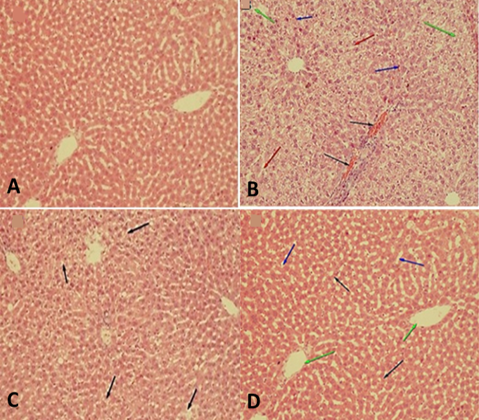
Histopathalogical changes in liver of animals induced by Triton X-100. Haematoxylin and Eosin (H and E) ×100. A: normal configurtion of liver. B: untreated Triton X-100 induced hyperlipidaemia rats having severe congestion of hepatic blood vessels (arrows). C: Simvastatin treated rats with mild fatty changes in hepatocytes. D: C. lanatus seeds treated rats having mild congestion of hepatic blood vessels.
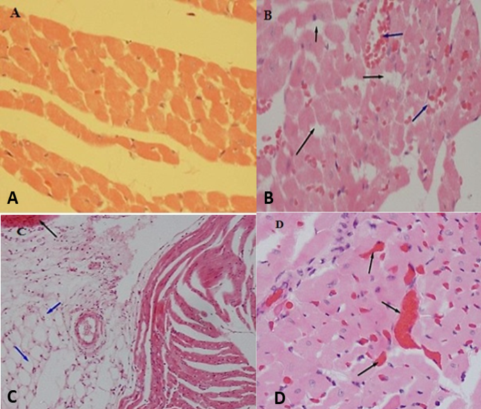
Effect of methanol extract of the plant seeds on histopathalogical changes in transverse section of hearts of rats induced by Triton X-100. A: normal configuration of heart muscle and coronary vessels. B: accumulation of lipid particles (black arrow) and severe congestion of blood coronary vessels (blue arrow). C: cardiac muscle fibres had vacuolation of lipids droplets (blue arrow) and mild congestion of coronary blood vessels (black arrow). D: mild congestion of blood coronary vessels with no hyperlipidaemia condition.
References
- Athyros, V.G., Tziomalos, K., Gossios, T.D., Griva, T., Anagnostis, P., Kargiotis, K. and GREACE Study Collaborative Group. 2010. Safety and efficacy of long-term statin treatment for cardiovascular events in patients with coronary heart disease and abnormal liver tests in the Greek Atorvastatin and Coronary Heart Disease Evaluation (GREACE) Study: a post-hoc analysis. Lancet, 376: 1916-1922. https://doi.org/10.1016/S0140-6736(10)61272-X[CrossRef]
- Calixto, J.B., 2005. Twenty-five years of research on medicinal plants in Latin America: A personal view. J. Ethnopharmacol., 100: 131-134. https://doi.org/10.1016/j.jep.2005.06.004[CrossRef]
- Carleton, H.M., Drury, R.A.B. and Wallington, E.A., 1980. Carleton’s histological technique. Oxford University Press. https://catalogue.nla.gov.au/Record/2864951
- Catapano, A.L., Farnier, M., Foody, J.M., Toth, P.P., Tomassini, J.E., Brudi, P. and Tershakovec, A.M., 2014. Combination therapy in dyslipidemia: Where are we now? Atherosclerosis, 237: 319-335. https://www.sciencedirect.com/science/article/pii/S0021915014014282, https://doi.org/10.1016/j.atherosclerosis.2014.09.026[CrossRef]
- Davidson, M.H., Voogt, J., Luchoomun, J., Decaris, J., Killion, S., Boban, D. and Neese, R., 2013. Inhibition of intestinal cholesterol absorption with ezetimibe increases components of reverse cholesterol transport in humans. Atherosclerosis, 230: 322-329. https://doi.org/10.1016/j.atherosclerosis.2013.08.006[CrossRef]
- Friedewald, W.T., Levy, R.I. and Fredrickson, D.S., 1972. Estimation of the concentration of low-density lipoprotein cholesterol in plasma, without use of the preparative ultracentrifuge. Clin. Chem., 18: 499-502. https://doi.org/10.1093/clinchem/18.6.499[CrossRef]
- Gramza, A. and Korczak, J., 2005. Tea constituents (Camellia sinensis L.) as antioxidants in lipid systems. Trends Fd. Sci. Tech., 16: 351-358. https://doi.org/10.1016/j.tifs.2005.02.004[CrossRef]
- Hassan, L.E.A., Koko, W.S., Osman, E.E., Dahab, M.M. and Sirat, H.M., 2011. In vitro antigiardial activity of Citrullus lanatus var. citroides extracts and cucurbitacins isolated compounds. J. med. Pl. Res., 5: 3338-3346. http://www.academicjournals.org/JMPR
- Hong, M.Y., Hartig, N., Kaufman, K., Hooshmand, S., Figueroa, A. and Kern, M., 2015. Watermelon consumption improves inflammation and antioxidant capacity in rats fed an atherogenic diet. Nutri. Res., 35: 251-258. https://doi.org/10.1016/j.nutres.2014.12.005[CrossRef]
- Koba, S. and Hirano, T., 2011. Dyslipidemia and atherosclerosis. Nihon Rinsho. Jpn. J. clin. Med., 69: 138-143. https://europepmc.org/article/med/21226274
- Kumar, A., Mazumder, A. and Saravanan, V., 2008. Antihyperlipidemic activity of Camellia sinensis leaves in Triton WR-1339 induced albino rats. Pharmacogn. Mag., 4: 60. https://www.researchgate.net/publication/333603466_Pharmacognosy_Magazine
- Li, H., Horke, S. and Förstermann, U., 2014. Vascular oxidative stress, nitric oxide and atherosclerosis. Atherosclerosis, 237: 208-219. https://doi.org/10.1016/j.atherosclerosis.2014.09.001[CrossRef]
- Lucky, O.O., John, U.O., Kate, I.E., Peter, O.O. and Jude, O.E., 2012. Quantitative determination, metal analysis and antiulcer evaluation of methanol seeds extract of Citrullus lanatus Thunb (Cucurbitaceae) in rats. Asian Pac. J. trop. Biomed., 2: S1261-S1265. https://doi.org/10.1016/S2221-1691(12)60396-7[CrossRef]
- Mendis, S., Puska, P., Norrving, B. and World Health Organization, 2011. Global atlas on cardiovascular disease prevention and control, WHO, Geneva.
- OECD, 2008. OECD guidelines for the testing of chemicals, Test 425. OECD Publishing, Paris.
- Omoboyowa, D.A. and Ajayi, A.A., 2016. Effect of methanolic extract of citrulus lanatus seeds on lipid profile and oxidative stress in acetaminophen intoxicated rats. Adv. Biomed. Pharm., 3: 87-93. https://doi.org/10.19046/abp.v03i02.02[CrossRef]
- Poduri, A., Rateri, D.L., Saha, S.K., Saha, S. and Daugherty, A., 2013. Citrullus lanatus ‘sentinel’(watermelon) extract reduces atherosclerosis in LDL receptor-deficient mice. J. Nutri. Biochem., 24: 882-886. https://doi.org/10.1016/j.jnutbio.2012.05.011[CrossRef]
- Ridker, P.M., Rifai, N., Cook, N.R., Bradwin, G. and Buring, J.E., 2005. Non–HDL cholesterol, apolipoproteins AI and B100, standard lipid measures, lipid ratiosand CRP as risk factors for cardiovascular disease in women. J. Am. med. Assoc., 294: 326-333. https://jamanetwork.com/, https://doi.org/10.1001/jama.294.3.326[CrossRef]
- Saravanan, R., Rajendra Prasad, N. and Pugalendi, K.V., 2003. Effect of piper betle leaf extract on alcoholic toxicity in the rat brain. J. med. Fd., 6: 261-265. https://doi.org/10.1089/10966200360716689[CrossRef]
- Sathya, J. and Shoba, F.G., 2014. Assessment of antimicrobial efficacy of Citrullus lanatus methanolic seed extract. J. chem. pharm. Res., 6: 640-643. https://pdfs.semanticscholar.org/d781/ b57cd2744b301e7bfc6d526cadc2cc5c008e.pdf
- Sharma, S., Sarvesh, P., Dwivedi, J. and Amita, T., 2011. First report on laxative activity of Citrullus lanatus. Pharmacologyonline, 2: 790-797. http://www.phytopharmajournal.com/V2issue606.pdf
- Shinde, S., Chivate, N., Kulkarni, P. and Naikwade, N., 2013. Hypolipidemic activity of Psidium guajava Linn leaves extracts in hyperlipidemic rats. Int. J. Pharm. Pharm. Sci., 5: 70-72. https://pdfs.semanticscholar.org/7b1a/a5d4c218bdd7fa79cf6c068fa909a1dbbb5c.pdf
- Singh Gill, N., Sood, S., Muthuraman, A., Bali, M. and Dev Sharma, P., 2011. Evaluation of antioxidant and anti-ulcerative potential of Citrullus lanatus seed extract in rats. Lat. Am. J. Pharm., pp. 30. http://sedici.unlp.edu.ar/handle/10915/8158
- Sudha, S.S., Karthic, R. and Naveen, J.R., 2011. Anti hyperlipidemic activity of Spirulina platensis in Triton x-100 induced hyperlipidemic rats. Hygeia J. D. Med., 3: 32-7. http://www.hygeiajournal.com/downloads/2337269785.sudha.pdf
- Sudhop, T., Lütjohann, D., Kodal, A., Igel, M., Tribble, D.L., Shah, S. and Von Bergmann, K., 2002. Inhibition of intestinal cholesterol absorption by ezetimibe in humans. Circulation, 106: 1943-1948. https://www.ahajournals.org/doi/abs/10.1161/01.cir.0000034044.95911.dc, https://doi.org/10.1161/01.CIR.0000034044.95911.DC[CrossRef]
- Terunuma, S., Kumata, N. and Osada, K., 2013. Ezetimibe impairs uptake of dietary cholesterol oxidation products and reduces alterations in hepatic cholesterol metabolism and antioxidant function in rats. Lipids, 48: 587-595. https://link.springer.com/article/10.1007/s11745-013-3790-6, https://doi.org/10.1007/s11745-013-3790-6[CrossRef]
- Valiathan, M.S., 1998. Healing plants. Curr. Sci., 75: 1122-1127. https://scholar.google.com.pk/citations?user=tyV77i8AAAAJandhl=enandoi=sra
- Winsor, L., 1998. Collection, handling, fixation, histological and storage procedures for taxonomic studies of terrestrial flatworms (Tricladida: Terricola). Pedobiologia, 42: 405-411. https://researchonline.jcu.edu.au/2715s
Services
Article usage statistics
Loading…