Anesthesia Consideration in a Dog with Degenerative Valve Disease Undergoing Soft Tissue Surgery
· · ·
Affiliations & Notes ⌄
1Kasetsart University Veterinary Teaching Hospital, Faculty of Veterinary Medicine, Kasetsart University, Kamphaeng Saen, Nakorn Pathom, Thailand
2Department of Large Animal and Wildlife Clinical Science, Faculty of Veterinary Medicine, Kasetsart University, Kamphaeng Saen Nakorn Pathom, Thailand. *Correspondence | Soontaree Petchdee, Department of Large Animal and Wildlife Clinical Science, Faculty of Veterinary Medicine, Kasetsart University, Kamphaeng Saen Nakorn Pathom, Thailand
Our study reported the techniques for anesthetic management in dogs with moderate to severe valvular degeneration during general anesthesia for soft tissue surgery.
This report suggests that the anesthetic drug protocol of diazepam plus alfaxalone and a maintenance fluid rate of 5 mL/kg/hr for dogs with valve disease is safe and appropriate for preserving cardiovascular function.
Selected from the article's own text.
Abstract
This case report aims to assist veterinarians in selecting appropriate anesthetic protocols and managing dogs with moderate to severe valvular degeneration during general soft tissue surgery. Three client-owned dogs with valve degeneration stages C or D who underwent general anesthesia for surgical treatment were included in this study. All dogs in this study were assigned to receive diazepam 0.3 mg/kg body weight, alfaxalone 2.5 mg/kg body weight, and a perioperative fluid rate of 5 ml/kg/hr. An appropriate assessment before and during surgery may help to prevent adverse events and preserve cardiovascular functions. This case report provides recommendations for anesthetic protocols and management in dogs with valvular heart disease. This study suggested that the anesthetic drug protocol of alfaxalone 2.5 mg/kg and a maintenance fluid rate of 5 mL/kg/hr in dogs with valve disease is safe and appropriate for preserving cardiovascular functions.
Keywords | Dog, General anesthesia, Mitral valve degeneration, Noncardiac surgery
Introduction
Degenerative heart valve disease (DVD) is the most common heart disease in ageing small-breed dogs . With the increasing number of dogs with DVD requiring surgical treatment, an appreciation of the impact of surgical and anesthetic treatments in the care of heart disease is essential. Previous studies have found that the risk of anesthetic death and the incidence of anesthetic complications in dogs increases with age . Surgical treatment can induce cardiac dysfunction, and dogs with heart valve disease who are undergoing surgery are often considered to be at higher risk of mortality and morbidity. However, proper care planning and the management of anesthetic drugs may help reduce this risk. An important aspect of anesthesia in dogs with valvular heart disease is the choice of anesthesia drugs, which may affect cardiac function, as well as the rate of fluid delivery during anesthesia. Anesthetic drugs that are commonly used in veterinary medicine include xylazine, ketamine, and propofol . Previous studies have reported anesthesia-related morbidity and mortality with the use of xylazine and isoflurane . Recent studies have shown toxic effects such as inducing cell injury and inflammation of the intravenous anesthetic propofol which is frequently used in dogs . Alfaxalone is a neuroactive steroid drug that has been confirmed for the induction or maintenance of anesthesia in dogs and cats in several countries over the past few years. A previous study showed that alfaxalone preserves cardiovascular functions and is safe for surgical use in dogs ; . Therefore, this case report presents a specific anesthesia protocol using alfaxalone and provides guidelines including fluid management during general anesthesia for dogs with heart valve disease.
Case History
The present case report describes three surgical cases in dogs with moderate to severe valve regurgitation (Table 1). The mean age and weight were 16±0.6 years and 4.2±0.8 kilograms, respectively. All dogs were categorized according to the American Society of Anesthesiologists (ASA) physical status 3.
Table 1: The table contains the breed, gender, age, weight, and DVD stage of the dogs included in the study.
Case 1
Case 2
Case 3
Breed
Crossbred
Poodle
Pomeranian
Age (year)
17
15
16
Gender
Male
Male
Female
Body weight (kg)
4.9
5.1
2.6
DVD stage
C
C
D
Case 1: A 17-year-old male crossbred dog weighing 4.9 kg presented to Kasetsart Veterinary Teaching Hospital Kamphaengsaen for resection of a subcutaneous mandibular mass. The dog had a history of degenerative mitral valve disease stage C. A mass was attached to the skin without adherence to the other tissues and the mass continued to grow over the course of 1 month.
Case 2: A 15-year-old male Poodle weighing 5.1 kg presented with periodontal disease. The dog had a history of degenerative mitral valve disease stage C.
Case 3: A 16-year-old female Pomeranian weighing 2.6 kg presented to Kasetsart Veterinary Teaching Hospital Kamphaengsaen for the surgical repair of an inguinal hernia. The dog had a history of degenerative tricuspid and mitral valve degeneration stage D.
Clinical evaluation and management
Dog owners were provided a consent form to approve the therapeutic procedure. A pre-anesthesia evaluation was performed, which comprised of routine history and physical examination, and laboratory parameters, including hematology, serum biochemistry, and electrolyte level analyses as shown in Table 2. A full cardiac examination was performed before and after the operation as shown in Tables 3 and 4.
Table 2: Blood profiles of three dogs.
Reference range
Case 1
Case 2
Case 3
HCT (%)
30-35
41.1
43.6
47.8
RBC (x106/µl)
5-9
6.34
6.13
7.03
HGB (gm%)
12-18
15.8
15.3
17.5
WBC (x103 µl)
6-17
12.73
14.19
12.08
Segmented
neutrophil (x103 µl)
3-11.5
69
79
72
Lymphocyte (x103 µl)
1-4.8
12
13
20
Monocyte (x103 µl)
0.15-1.35
6
5
5
Eosinophil (x103 µl)
0.1-1.25
13
3
3
Platelet (x103 µl)
200-900
300
200
400
BUN (mg%)
15-34
22.9
51.8
55
Creatinine (mg%)
<1.8
0.68
1.19
0.84
Total protein (gm%)
5.3-7.8
7.6
8.4
7.2
ALT (U/L)
28-74
80
118
47
HCT = hematocrit; RBC = Red blood cell; HGB = hemoglobin; WBC = White blood cell; BUN = Blood urea nitrogen; ALT = Alanine aminotranferase
Anesthesia techniques
The goal of anesthesia is to administer a combination of short-acting medications to provide sedation and analgesia while maintaining spontaneous respiration and airway reflexes. In this study, total intravenous anesthesia was administered with a continuous infusion of alfaxalone to
Table 3: Echocardiography parameters of three dogs.
Parameters
Case 1
Case 2
Case 3
Before
After
Before
After
Before
After
IVSd (cm)
0.6
0.6
0.6
0.6
0.6
0.6
LVIDd (cm)
3.0
3.0
3.3
3.2
1.3
1.2
LVPWd (cm)
0.5
0.5
0.6
0.6
0.4
0.5
IVSs (cm)
0.6
0.6
0.7
0.7
0.7
0.6
LVIDs (cm)
2.1
2.0
2.3
2.3
0.8
0.7
LVPWs (cm)
0.6
0.7
0.7
0.8
0.7
0.7
FS (%)
30
33
30
28
38
41
LA Diameter (cm)
2.0
1.9
2.4
2.5
1.9
1.5
AO Diameter (cm)
1.1
1.1
1.4
1.4
1.0
1.0
LA: AO
1.82
1.91
1.69
1.78
1.96
1.45
MV E Vel (m/s)
0.53
0.48
0.58
0.49
0.48
0.35
MV A Vel (m/s)
0.63
0.65
0.83
0.98
0.66
0.75
MV E/A ratio
0.85
0.73
0.70
0.5
0.73
0.48
TV E Vel (m/s)
0.29
0.48
0.56
0.7
0.5
0.65
TV A Vel (m/s)
0.36
0.58
0.93
0.98
0.75
0.93
TV E/A ratio
0.80
0.82
0.60
0.71
0.66
0.70
IVRT (ms)
50
55
67
75
41
45
TAPSE (mm)
120
110
130
120
100
90
IVSd; diastolic interventricular septum thickness, IVSs; systolic interventricular septum thickness, LVIDd; left ventricular end diastolic diameter, LVIDs; left ventricular end systolic diameter, LVPWd; left ventricular wall diastolic thickness, and LVPWs; left ventricular wall systolic thickness. FS = fractional shortening; IVRT = isovolumic relaxation time; TAPSE=Tricuspid annulus plane systolic excursion
Table 4: The respiratory and cardiovascular parameters of three dogs.
Parameters
Case 1
Case 2
Case 3
Before
After
Before
After
Before
After
Respiratory rate (bpm)
40
25
50
40
40
30
Heart rate (bpm)
120
140
176
180
119
137
SDNN
45
47
90
94
63
68
LF/HF ratio
0.63
0.78
0.46
0.60
1.2
1.5
MAP (mmHg)
130
140
70
75
75
80
SDNN=standard deviation of the R-R intervals, LF/HF= low frequency per high frequency ratio, MAP= mean arterial blood pressure.
gether with an opioid. All dogs were premedicated with an intravenous bolus of diazepam (0.3 mg/kg). General anesthesia was induced intravenously with alfaxalone. (2.5 mg/kg) and was maintained with isoflurane in 100% oxygen (3 L/min). Anesthesia monitoring was performed using pulse oximetry, electrocardiography, spirometry, and body temperature.
Thoracic radiography
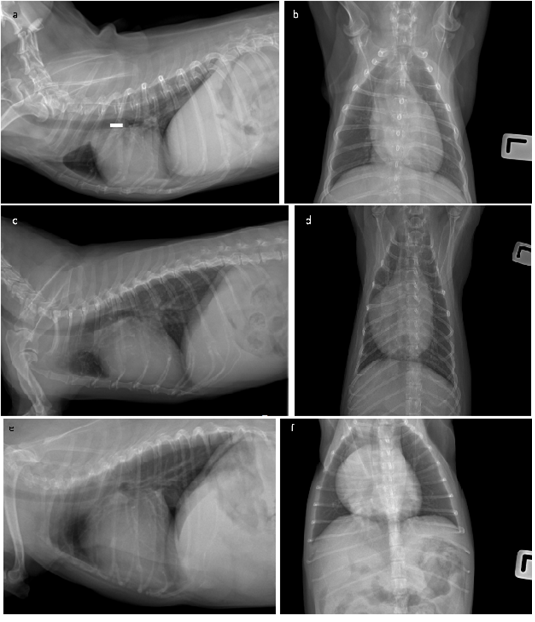
Cardiomegaly was diagnosed on chest radiography in all three cases. A thoracic radiograph is shown in Figure 1.
Echocardiography and heart rate variability measurement
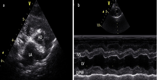
The severity of valve regurgitation was determined noninvasively via echocardiography (vivid s5, USA). Echocardiography was performed in parasternal long and short axis views and apical four-chamber views in the right and left parasternal positions with no sedation. Echocardiographic images were captured and stored for offline analysis. The left atrium dimension was measured by a standard left atrium to aorta ratio (LA/Ao). Left ventricular wall structure and function were calculated by measuring the images from two-dimensional and M-mode planes (Figure 2). Heart rate variability was measured by placing five electr--odes on the skin of the thorax to provide the ECG recordings (BTL Medical Technologies, Thailand).
Results and Discussion
Our study reported the techniques for anesthetic management in dogs with moderate to severe valvular degeneration during general anesthesia for soft tissue surgery. In all three cases included in this study, the advanced stages of degenerative valve disease and ventricular systolic, and diastolic dysfunction were presented and were typically masked by eccentric hypertrophy along with a markedly dilated left atrium. The left ventricle became less compliant, and the filling pressures increased. Measuring only the left ventricular functions, such as fractional shortening, may underestimate the severity of myocardial dysfunction. Dogs with chronic valve disease can experience pulmonary vascular changes, pulmonary hypertension, and right ventricular impairment. The tricuspid annular plane systolic excursion (TAPSE) is well correlated with the right heart function. A previous study reported that TAPSE <110 mm showed right ventricle dilation at the basal level and a worse degree of tricuspid regurgitation. In addition, isovolumic relaxation time (IVRT) and the E/A ratio are important echocardiography parameters for indicating an impairment of diastolic function . Not only the severity of valve regurgitation but also heart function, such as contraction and relaxation, must be considered. In addition, dogs with valvular heart disease with symptoms such as dyspnea, should undergo an anesthetic risk evaluation and consider a more detailed preoperative evaluation such as a chest X-ray and full scan echocardiography should be considered to reduce the potential risk of respiratory failure during general anesthesia.
Moreover, dogs should be treated with afterload reduction, diuretics, and inotropic support before surgery. Angiotensin-converting enzyme (ACE) inhibitors are frequently administered to dogs with chronic heart valve regurgitation. However, ACE inhibitors have been reported to be associated with perioperative hypotension. Thus, the ACE inhibitors should be withdrawn perioperatively, and continuous blood pressure monitoring should be considered for dogs with impaired myocardial function and presenting signs of congestive heart failure. Inotropic agents with β-1 agonistic effects, such as dobutamine or epinephrine should be prepared in cases of systemic hypotension.
The general anesthetic goals are to promote forward flow by maintaining a high heart rate and less afterload. The results of this study are similar to a previous report that the anesthetic protocol using diazepam and alfaxalone produced an increase in HR and influenced sympathetic activity (Pattanapon., 2018). These results suggested that diazepam and alfaxalone might preserve the cardiac autonomic activity balance. Using fluid maintenance during anesthesia can cause hypervolemia complications in patients with cardiovascular disease. An initial crystalloid fluid rate of 5 mL/kg/hr is recommended for healthy dogs undergoing anesthesia . In this study, we recommended providing a maintenance rate of 5 mL/kg/hr for dogs with heart valve disease and undergoing general anesthesia for soft tissue surgery. However, therapy must be individualized and adjusted to each dog.
Conclusions and recommendations
Previous studies have reported the risk of operative morbidity and mortality in patients with cardiovascular diseases. This report suggests that the anesthetic drug protocol of diazepam plus alfaxalone and a maintenance fluid rate of 5 mL/kg/hr for dogs with valve disease is safe and appropriate for preserving cardiovascular function.
Acknowledgments
The authors are grateful to the owner and Kasetsart Veterinary Teaching Hospital Kamphaeng Saen, Thailand for providing the necessary facilities.
Novelty statement
The novelty of this case report is the guidance protocol to assist veterinarians in selecting appropriate anesthetic drugs and managing dogs with heart disease.
Conflict of interest
The authors declare that there is no conflict of interest.
Author’s contribution
All the authors contributed to the manuscript. Chattida Panprom, Wannisa Meepoo, and Nakrob Pattanapon prepared and interpreted the results and Soontaree Petchdee drafted, critically revised, and approved a manuscript.
Figures
Figure 1. Figure 1.
Radiograph of thoracic on lateral and dorsoventral view in three cases.
Figure 2.
Echocardiographic of the left atrium and aorta ratio (a) and M mode image of the left ventricle of dog with valve degeneration (b). AO= Aorta, LA = left atrium, and LV=left ventricle.
References
Beiseigel M., Simon B.T., Michalak C., Stickney M.J., Jeffery U. (2021). Effect of peri-operative crystalloid fluid rate on circulating hyaluronan in healthy dogs: A pilot study. Vet. J. 267: https://doi:10.1016/j.tvjl.2020.105578[CrossRef]
Bosnjak Z.J., Logan S., Liu Y., Bai X. (2016). Recent Insights into Molecular Mechanisms of Propofol-Induced Developmental Neurotoxicity: Implications for the Protective Strategies. Anesthes. Analg. 123(5):1286-1296. https://doi.org/10.1213%2FANE.0000000000001544
Carter J., Story D.A. (2013). Veterinary and human anesthesia: an overview of some parallels and contrasts. Anesthes. Intens. Care. 41(6): 710-718. https://doi.org/10.1177/0310057x1304100605[CrossRef]
Chiu K.W., Robson S., Devi J.L., Woodward A., Whittem T. (2016). The cardiopulmonary effects and quality of anesthesia after induction with alfaxalone in 2-hydroxypropyl-β-cyclodextrin in dogs and cats: a systematic review. J. Vet. Pharmacol. Therapeut. 39(6): 525-538. https://doi.org/10.1111/jvp.12312[CrossRef]
Davis H., Jensen T., Johnson A., Knowles P., Meyer R., Rucinsky R., Shafford H. (2013). AAHA/AAFP Fluid therapy guidelines for dogs and cats. J. American Anim. Hosp. Assoc. 49:149-159. https://doi.org/10.5326/jaaha-ms-5868[CrossRef]
Dyson D.H., Maxie M.G., Schnurr D. (1998). Morbidity and mortality associated with anesthetic management in small animal veterinary practice in Ontario. J. American Anim. Hosp. Assoc. 34(4): 325-335. https://doi.org/10.5326/15473317-34-4-325[CrossRef]
Mattin M.J., Boswood A., Church D.B., López-Alvarez J., McGreevy P.D., O’Neill D.G., Thomson P.C., Brodbelt D.C. (2015). Prevalence of and risk factors for degenerative mitral valve disease in dogs attending primary-care veterinary practices in England. J. Vet. Inter. Med. 29(3): 847-854. https://doi.org/10.1111%2Fjvim.12591
Matthews N.S., Mohn T.J., Yang M., Spofford N., Marsh A., Faunt K., Lund E.M., Lefebvre S.L. (2017). Factors associated with anesthetic-related death in dogs and cats in primary care veterinary hospitals. J. American Vet. Med. Assoc. 250(6): 655-665. https://doi.org/10.2460/javma.250.6.655[CrossRef]
Pattanapon N., Bootcha R., Petchdee S. (2018). The effects of anesthetic drug choice on heart rate variability in dogs. J. Adv. Vet. Anim. Res. 5(4): 485-489. https://doi.org/10.5455%2Fjavar.2018.e303
Petchdee S., Yalong M., Kaewnet M., Ithisariyanont B., Padawong T. (2022). Assessment of Right Ventricular Function, Blood Lactate Levels, and Serum Peptidomics Profiles Associated with Mitral Valve Disease in Dogs. Frontier Vet. Sci. 20: 8:789137. https://doi.org/10.3389/fvets.2021.789137[CrossRef]