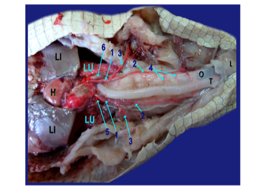
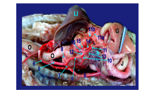
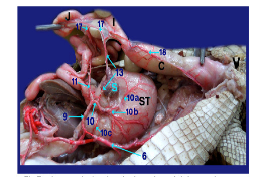
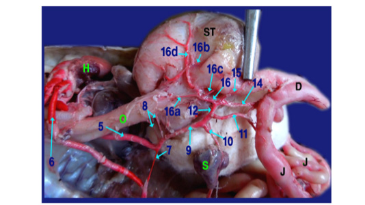
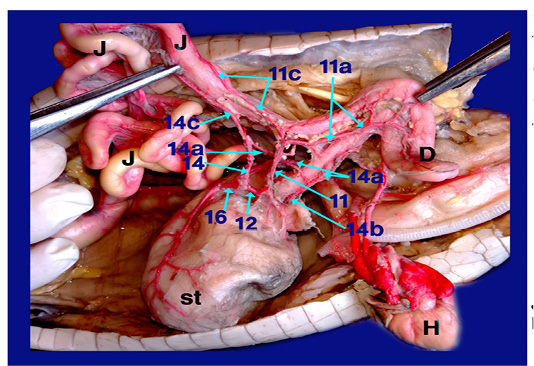
1)Brachicephalic A., 2)Common Carotid A., 3)Subclavian A., 4)Esophageal branches of common carotid A., 5)Left aorta, 6)Right aorta, 7)Short communicating branch, 8)Branches to the terminal part of esophagus, 9)Celiacomesentric A., 10)Right gastric A., 10a)Branch to cardiac region of stomach, 10b)Branch to body of stomach, 10c)Branch to caudal end of stomach, 11)Intestinal A., 11a) Branch to1st part of duodenum and outer loop, 11b) Hepatic branch, 11c)Dorsal jejunal branch, 12)gastroduodenal A., 13)Splenic A., 14)duodenal A., 14a) Branch supplied the 2nd loop of duodenum., 14b)Fine branches of duodenal artery to the small sac of stomach, 14c) branches to ventral wall of initial part of jejunum 15)Pyloric A., 16)left gastric A., 16a) branches of left gastric artery to cardiac region, 16b) branches of left gastric artery to small portion of stomach, 16c) branches of left gastric artery to pyloric region, 16d) Twigs of left gastric artery to the body of stomach, 17) Jejunal A., 18) Colic branches.
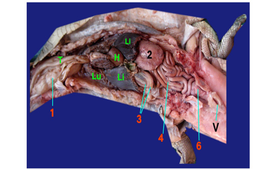
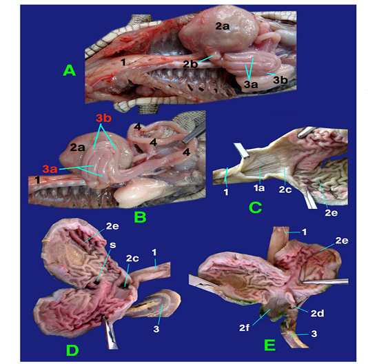
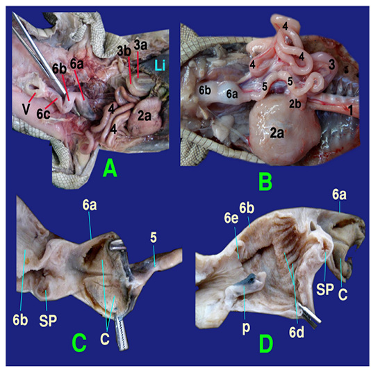
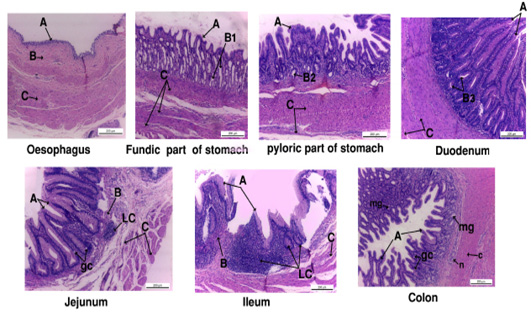
H) Heart, Lu)Lung, T)Trachea, L)Larynx, O)Esophagus, St)Stomach, D)Duodenum, J)Jejunum, C)Colorectum, V)Vent, S)spleen, Li)Liver, G)Gonad.
The Right gastric artery gave splenic artery (fig.6, 7/13) then split into three main branches on right face of stomach; branch to the cardiac region, one to the body and other to the terminal caudal end of stomach (fig. 7 /10a,10b and 10c respectively).
The intestinal A. supplied 1st part of duodenum (fig. 6, 9/11a) with its second loop (outer loop) included pancreas and gave hepatic branches (fig. 6/11b) to right and left lobe of liver. It ran on the dorsal wall of jejunum (1st dilated part about 4cm) as dorsal jejunal branch (fig.9/11c).
The gastro duodenal artery gets out duodenal A., Pyloric A. and left gastric A. (fig. 6, 8, 9 /14, 15 and 16 respectively).
A) Duodenal A. (the largest branch) (fig. 6,8,9 /14) supplied the 2nd loop of duodenum (inner loop) (fig.9/14a), pancreas and gave several fine branches to the small sac of stomach (fig. 9/14b). It also gave ventral jejunal branch (fig. 9 /14c) to the ventral wall of initial part of jejunum.
B) Pyloric A. (fig. 6, 8 /15) supplied the terminal part of stomach (pyloric region) then continued to the right lobe of liver.
C) Left gastric A. (fig. 6, 8, 9 /16) passed to the left face of stomach giving several fine branches (fig. 8 /16a) to cardiac region, small portion of stomach (fig. 8 /16b) and pyloric region (fig. 8/16c), also gave few twigs (fig. 8 /16d) to the body of stomach.
Right aorta (fig. 6, 7, 8/6) gave jejunal A. (fig. 7/17) which divided into 5-6 primary jejunal branches which gave secondary jejunal branches and finally terminated as intestinal arches.
Splenic artery (fig. 6, 7/13) was a branch from the right gastric artery which gave proper splenic branch in its hilus then anastomosed with last jejunal artery supplying the terminal part of jujenum (ileum before change in diameter from narrow to wide) then passed on the dorsal aspect of colon giving colic branches (fig. 7 /18) to supply the dorsal aspect of colorectum till the cloaca.