Abstract | Among helminthes, the trichostrongyloidea consists of most significant parasites of ruminants, which are posing significant threats to livestock productivity, health and well-being.
Editor | Muhammad Imran Rashid, Department of Parasitology, University of Veterinary and Animal Sciences, Lahore, Pakistan.
Gastrointestinal nematode infections are plentiful and among the most expensive illnesses in the domesticated animals industry.
Anthelmintic resistance in nematode parasites of cattle: a global issue?
1Department of Parasitology, Gomal University Dera Ismail Khan, Khyber Pakhtoon Khah, Pakistan; 2Department of Parasitology, University of Veterinary and Animal Sciences Lahore, Punjab, Pakistan; 3Quality Operations Laboratory, University of Veterinary and Animal Sciences, Lahore, Pakistan; 4University of Edinburgh, Royal (Dick) School of Veterinary Studies and Roslin Institute,Easter Bush Veterinary Centre, Midlothian, Scotland, EH25 9RG, UK.
Abstract | Among helminthes, the trichostrongyloidea consists of most significant parasites of ruminants, which are posing significant threats to livestock productivity, health and well-being. This brief commentary provides an overview of this important group of parasite to showcase the crucial roles these play in hosts.
Classification and Life cycle of the Trichostorngyloidea
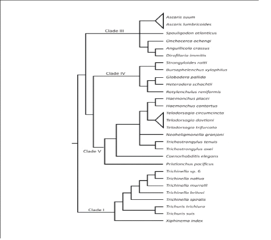
The phylum Nemathelminths has six branches, the nematodes form one of them, which contains parasite of veterinary importance.Nematodes are ordinarily called roundworms, due to their look of rounded body structure. These roundworms are additionally subdivided into order, superfamily, genus, species and a most essential divisions: bursate and non-bursate clusters due to the presence of bursa . Mostly individuals from the order Strongylida of nematoda class bursate group, which additionally subdivided into 4 diverse superfamilies: the trichostrongyloidea (contain most significant parasites of ruminants), the strongyloidea (contain numerous significant parasites of equine),the ancylostomatidae the (contain hookworms of the human and pets) and the metastrongyloidea (contain worms of lungs of pets animals) . After the approach of molecular genetics, the evolutionary system has been built up on the basis of little subunit of DNA of the ribosomes which separate the parasitic roundworms hereditarily into five important clades I-V (Figure 2.1) . The roundworms in the superfamily trichostongyloidea (Clade V) which are most essentialgastrointestinal (GI) parasites of ruminant livestock animals . The major GI nematode types of cattle in the superfamily trichostongyloidea are Haemonchusplacei, O.ostertagi, M.digitatus and T.axei present in the abomasum; S.papillosus,Cooperiaand Nematodirrus parasites are present in the small intestinal tract; and O.radiatum present in large intestinal tract ; . Among every single above species,H. placei, C. oncophora and O.ostertagi are estimated the most predominant in bovine; especially the rate of infection of H. placei was very high in cattle.
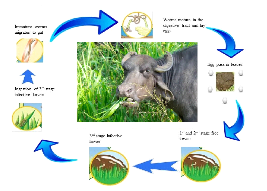
The life cycles of roundworm parasites are direct and extensively same in all parasites (see in Figure 2.1). On account of gastrointestinal roundworms, the stages of growth occur in the GI tract, where they lay extensive quantities of eggs by the fertilized females that are released from body through feces. Trichostrongyloidea eggs are incubate done to two days inside the dung and hatched into L1 stage larvae, which are further, transformed into the L2 and L3 stage larvae inside the dung. The L3 is the infective stage larvae, which relocate their position from dung to pasture, fodder, water reservoir and once L3ingested by the animal, molt into L4 larvae. After thatL4 is converted into adult in the GI tract. After mating with male, the adult female worms begin laying eggs at day 18.An amazing egg production of a fertilized female reaches to range between 5000– 15,000 eggs per day (Figure 2.2) .
• Anderson TJC, Blouin MS, Beech RN (1998). Population Biology of Parasitic Nematodes: Applications of Genetic Markers, In: Baker JR, Muller R, Rollinson D. (Eds.) Advances in Parasitology. Academic Press, pp. 219-283. https://doi.org/10.1016/S0065-308X(08)60425-X[CrossRef]
• Chan MS (1997). The global burden of intestinal nematode infections — Fifty years on. Parasitol. Today. 13: 438-443. https://doi.org/10.1016/S0169-4758(97)01144-7[CrossRef]
• Fox MT (1997). Pathophysiology of infection with gastrointestinal nematodes in domestic ruminants: recent developments. Veterinary parasitology 72: 285-297. discussion 297-308. https://doi.org/10.1016/S0304-4017(97)00102-7[CrossRef]
• Fukumoto Si, Etani K, Toi K, Hanadate M, Hidaka M, Yokoya K, Hiramatsu T, Iguchi T, Kudo S, Miyamoto K, Bando G (1990). Epidemiology of Abomasal Nematodes of Dairy Cattle in Hokkaido, Northern Japan. Japanese J. Vet. Sci. 52: 379-385. https://doi.org/10.1292/jvms1939.52.379[CrossRef]
• Gilabert A, Wasmuth JD (2013). Unravelling parasitic nematode natural history using population genetics. Trends Parasitol. 29: 438-448. https://doi.org/10.1016/j.pt.2013.07.006[CrossRef]
• Gilleard JS (2013). Haemonchus contortus as a paradigm and model to study anthelmintic drug resistance. Parasitology. 140: 1506-1522. https://doi.org/10.1017/S0031182013001145[CrossRef]
• McLeod RS (1995). Costs of major parasites to the Australian livestock industries. Int. J. Parasitol. 25: 1363-1367. https://doi.org/10.1016/0020-7519(95)00071-9[CrossRef]
• Murrell KD (1991). Economic losses resulting from food-borne parasitic zoonoses. Southeast Asian J. Trop. Med. Pub. Health. 22 Suppl: 377-381.
• Oku Y, Nakazawa M, Hatakeyama S, Miyaji S, Kitaguchi T, Cabrera-Lopez CA, Okamoto M, Kamiya M, Ohbayashi M, Ooi HK (1987). A survey of abomasal and duodenal nematodes in cattle in Hokkaido, Japan. Japanese J. Vet. Res. 35: 67-72.
• Redman E, Whitelaw F, Tait A, Burgess C, Bartley Y, Skuce PJ, Jackson F, Gilleard JS (2015). The Emergence of Resistance to the Benzimidazole Anthlemintics in Parasitic Nematodes of Livestock Is Characterised by Multiple Independent Hard and Soft Selective Sweeps. PLoS Neglect. Trop. Dis. 9: e0003494. https://doi.org/10.1371/journal.pntd.0003494[CrossRef]
• Stepek G, Buttle DJ, Duce IR, Behnke JM (2006). Human gastrointestinal nematode infections: are new control methods required? Int. J. Experimen. Pathol. 87: 325-341. https://doi.org/10.1111/j.1365-2613.2006.00495.x[CrossRef]
• Stromberg BE, Gasbarre LC (2006). Gastrointestinal nematode control programs with an emphasis on cattle. Vet. Clin. North America. Food Anim. Pract. 22: 543-565. https://doi.org/10.1016/j.cvfa.2006.08.003[CrossRef]
• Sutherland IA, Leathwick DM (2011). Anthelmintic resistance in nematode parasites of cattle: a global issue? Trends Parasitol. 27: 176-181. https://doi.org/10.1016/j.pt.2010.11.008[CrossRef]
• Urquhart G, Armour J, Duncan J, Dunn A, Jennings F (1996). Veterinary helminthology. Vet. Parasitol. 2: 35-38.
• Zajac AM (2006). Gastrointestinal Nematodes of Small Ruminants: Life Cycle, Anthelmintics, and Diagnosis. Vet. Clin. Food Anim. Pract. 22: 529-541. https://doi.org/10.1016/j.cvfa.2006.07.006[CrossRef]
• Ziegelbauer K, Speich B, Mausezahl D, Bos R, Keiser J, Utzinger J (2012). Effect of sanitation on soil-transmitted helminth infection: systematic review and meta-analysis. PLoS Med. 9: e1001162. https://doi.org/10.1371/journal.pmed.1001162[CrossRef]