AD is a multifactorial neurodegenerative disease which involves different processes and difficult to manage.
The socioeconomic needs to overcome the progression of disease takes time.
Current drug therapy only allows the symptomatic relief by maintaining the level of acetylcholine in the brain while the neuronal death rate cannot be altered.
So, further consideration is needed for the development of medication for the disease.
Selected from the article's own text.
Abstract
Alzheimer’s disease (AD) is a degenerative disease of neurons in which brain cells started to loss their functions. Its main cause is dementia which results in decreased activity of brain such as thinking and increased dependence on others to fulfill the daily tasks. The pathological identification of AD is beta amyloid accumulation or development of neurofibrillary tangles of tau proteins. Currently different hypothesis was proposed in the progression of disease which are amyloid cascade, tau and cholinergic hypothesis. Other than that age, family history, injury, high blood pressure and genetic may play an important role in the disease development. Current AD treatment are acetylcholinesterase inhibitors are easily accessible in the market to relief the AD primary symptoms but did not cure it. AD treatment needs a therapeutic approach to minimize its progression. This review focuses on the understanding of disease, hypothesis and treatment of disease to slow its progression.
Novelty Statement | Amidst the challenges posed by the current medications, the narrative extends into the realm of experimental drug therapy, shedding light on the potential breakthroughs, such as aducanumab, while acknowledging the ongoing quest for more targeted treatments that address the fundamental causes of this multifactorial neurodegenerative disease.
Introduction
Alzheimer’s disease (AD) is a progressive degenerative disorder of brain characterized by the decline in cognitive and memory impairment due to amyloid plagues and neurofibrillary tangles which results in the loss of neurons . This includes the loss of recent and episodic memory, as well as difficulties with language, attention, problem-solving and decision-making ; . The AD symptoms typically worsen over time, leading to significant impairment in daily functioning (Varzaghani et al., 2022). Given that AD is a chronic, progressive illness, its prognosis is not entirely known. With the disease progression, individuals with AD may experience more severe symptoms like uncertainty, disorientation, agitation and behavioral and personality changes . In addition to cognitive symptoms, AD can also present with behavioral and psychological changes. These changes can include depression, anxiety, irritability, aggression, and psychosis .
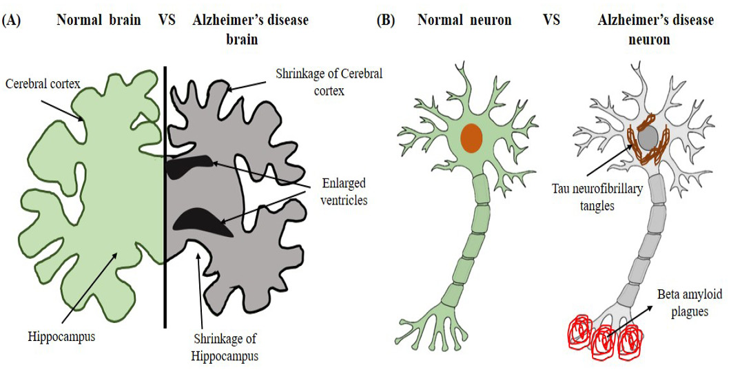
A person with AD has brain alterations. It becomes smaller with time and has fewer healthy cells. β-amyloid plaques, which are clusters of protein that build up between the nerve cells rather than vanishing as they would in healthy brains and neurofibrillary tangles, which are twisted fibers inside brain cells that obstruct the movement of particles and other essential substances from one part of the cell to another, are two of the pathological signs of AD . The plagues and tangles in the brain harmed the surrounding brain cells . When damaged cells die, the cerebral cortex shrinks. These changes result in signs of AD, such as changes in mood, confusion, loss of memory, and speech difficulty (Figure 1A). Amyloid plaques have been seen in the autopsied brains of AD patients since Dr. Alois Alzheimer’s time, indicating that these diseases are what cause the illness .
A recent study conducted by World Health organization said that there are about fifty-five million people are living around the globe with dementia and AD . The AD population is divided into early or late onset of disease of which around two hundred thousand people younger than 65 years old and five million are above 65 years of age, later represents the late onset of disease . It is estimated that 13.8 million people will have AD by 2050, meaning that a new case will occur every 33 seconds, or more than a million new cases annually ; .
Causes of AD
Genetic, environmental and behavioral factors, together with age-related alterations in the brain, are probably important causes. Depending on the individual, each of these factors may or may not be as significant in raising or lowering the risk of AD .
Certain factors play an important part to increase the chance in person to developed AD. So far, researches have concluded that people with 65 or older age get the AD, gender plays an important role in AD occurrence that women are more pronounced to AD then men, if AD runs in family, then individual are more likely to developed AD, people with Down’s syndrome likely to have AD in their 30s or 40s. Other factors include head injury, high cholesterol, high blood pressure and lower level of acetyl cholinesterase ; .
Stages of AD
There are seven stages of AD through which it progresses to loss of neurons in brain and behavioral changes of individual. Stage I is no impairment stage which shows no physical symptoms of AD . At stage II, forgetfulness rises in AD person with no proper medication . Stage III and IV started to show AD signs of declined brain functioning such as reasoning and thinking . In stage V and VI, AD person become more agitated, required assistant in daily activities and confusion persist . Stage VII is the last stage of AD in which person ability to respond to environment become zero and he may lose the ability to move.
AD pathogenesis
AD is characterized by a specific pattern of pathological alterations in the brain. These changes include the formation of intracellular tau protein tangles and extracellular β-amyloid plaques, which causes neurodegeneration and loss of synaptic connections . The β-amyloid plaques are made up of clumps of β-amyloid proteins, is believed to contribute to the disruption of normal neural function and the initiation of a cascade that would lead to neural dysfunction and brain cell death . On the other side, aberrant tau protein phosphorylation results in the formation of tau protein tangles (Figure 1B). In order to preserve adequate cellular function and transport, neurons’ microtubule structures must not be damaged by this aberrant phosphorylation. Neurodegeneration, loss of synaptic connections, and increasing memory problems and cognitive impairment are the results of these degenerative alterations in the brain ; . While the exact pathophysiology of AD remains largely unknown, a combination of genetic, environmental, and lifestyle factors are believed to be involved. Although the fundamental aetiology of AD is not entirely understood, there are many ideas as to how it develops. According to some views, the development of tau protein tangles and β-amyloid plaques may be the main causes of AD pathogenesis. The onset and progression of Alzheimer’s disease (AD) have been linked to various variables, including inflammation and cerebrovascular disease, although the buildup of tau protein tangles and β-amyloid plaques are commonly acknowledged as the main pathological characteristics of the disease. There is strong consensus that AD pathology converges in the synapse, despite continuous disagreements. AD is primarily initiated and propagated by tau and β-amyloid protein clumps ; . While the exact mechanisms by which β-amyloid and tau pathology contribute to AD are still under investigation, it is widely accepted that these protein aggregates disrupt neuronal communication and lead to synaptic dysfunction, neurodegeneration, and cognitive decline. Research suggests that aberrant connections between cerebral regions involved in cognitive functioning contribute to the pathogenesis of AD. Furthermore, research has demonstrated that modifications to the epigenetic code, including as acetylation, methylation, and PARylation, may potentially play a role in the pathophysiology of AD by controlling gene expression and changing the function of proteins ; . Other theory propose that due to inflammation, oxidative stress, and mitochondrial dysfunction may also play a role in the development and progression of the disease by decreasing the level of brain cholinesterase ; ; . Some other researchers suggested the anti-oxidative potential Scrophularia amplexicaulis in inviro model of AD by increasing the cholinesterase activity . In the process of lipid peroxidation, many free radicals formed which damage the cell membranes . Enzymes like catalase and superoxide dismutase counteract this damage ; . Catalase breaks down harmful hydrogen peroxide, while superoxide dismutase converts superoxide radicals into less harmful molecules. Nitric oxide plays a dual role, acting as a signaling molecule and a potential source of oxidative damage when excessive . However, it is crucial in regulating the redox balance which emphasizes the importance of a balanced antioxidant defense system for cellular health.
However, early diagnosis and intervention can help to improve the living condition of the patient and slow disease progression. Prognosis of AD is very difficult as it is in progressive nature, which ultimately causes significant cognitive decline and functional impairment. This can result in a significant burden on caregivers and healthcare systems.
AD molecular mechanisms hypothesis
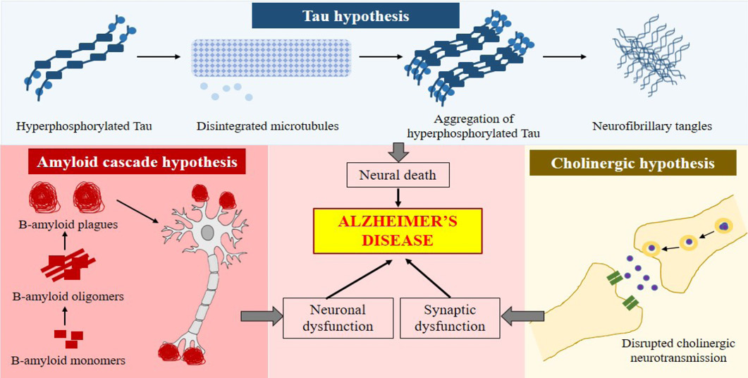
Different mechanisms occur during the progression of AD. Few of the hypothesis were presented in the progression of AD such as β-amyloid cascade, tau and cholinergic hypothesis. Schematic illustration of these hypothesis was presented in Figure 2.
Beta-amyloid cascade hypothesis
The β-amyloid cascade hypothesis is one of the predominant theories in AD research over the past two decades . According to the β-amyloid cascade hypothesis, the deposition of β-amyloid plaques is the first step in the pathogenesis of AD, followed by the cascade of inflammatory responses and formation of tau neurofibrillary tangles from hyper phosphorylated tau proteins . It also suggests that the abnormal accumulation and aggregation of β-amyloid peptide is the main causative agent of AD, which lead to subsequent neurodegeneration and cognitive decline . Some researchers argue that β-amyloid accumulation may be a consequence rather than a cause of disease. As a result, a mix of genetic, environmental, and lifestyle variables have a part in the etiology of AD, which is complicated and multifaceted. Many risk factors have been linked to a higher likelihood of acquiring AD, including pollution, diabetes, anoxia, high blood pressure, low educational attainment, and a sedentary lifestyle. The prognosis for individuals with AD is generally poor, as there is no complete cure for the condition.
Tau hypothesis
The tau hypothesis states that normal adult tau transforms into neurofibrillary tangles due to excessive or abnormal tau phosphorylation. Mutations affecting tau protein synthesis and function result in hyperphosphorylation in the brain ; . Although the cause of tau aggregation in the absence of mutations is unknown, hyperphosphorylated tau proteins, which deconstruct microtubules and entrap normal tau amino acids, may be a contributing factor . This irreversible structure impairs cytoplasmic processes and hinders axonal transport, both of which might result in cell death . Since the amyloid hypothesis is attracting more attention than tau over time, the tau hypothesis is frequently at an earlier stage. Nonetheless, tau-based strategies have shown some promising results, and phase II trials are currently being conducted on seven anti-tau medications .
Table 1: Management of AD through medicines.
Drugs
Initial dose
Maximum dose
Indications
Side effects
Donepezil
5mg orally once a day
23 mg once a
day
Mild to severe
Nausea, vomiting, diarrhea, insomnia, muscle cramps, fatigue and weight loss
Galantamine
4 mg orally twice a day
12 mg orally twice a day
Mild to moderate
Nausea, vomiting, diarrhea, decreased appetite, weight loss, dizziness and headache
Memantine
5 mg orally twice a day
10 mg orally twice a day
Moderate to severe
dizziness, headache, diarrhea, constipation, and confusion
AD is a chronic condition that causes memory loss as a result of cell death in the hippocampus of the brain, which is primarily regulated by cholinergic regulation . According to this theory, the cholinergic neuronal loss in the hippocampus is affected by changes in neurotransmitters level. Numerous researches were conducted on AD brain and it revealed lower levels of choline acetyltransferase, acetylcholine release, nicotine receptors and also muscarinic receptors in brain . The acetylcholinesterase inhibitors drug that is currently licensed for treating ADD (attention deficit disorder) and improving the uptake of acetylcholine by synapses. Unfortunately, none of these drugs have the ability to alter AD’s course or even considerably slow down how quickly the condition progresses ; . Their therapeutic effects are palliative. However, the possibility that they may be utilized in conjunction with other disease-modifying medications in therapy should not be discounted.
Current medication of AD
AD is a progressive degenerative disease of brain characterized by the decline in cognitive and memory impairment due to the loss of neurons. As the age of world population increases, effective drugs that can halt AD’s growth and enhance the lives of individuals who are afflicted are becoming more and more necessary. Currently, the medications available for AD provide only modest and transient benefits to a subset of patients. Acetylcholinesterase inhibitors and memantine are two commonly prescribed medications for patients with mild-moderate AD . Acetylcholinesterase inhibitors increased the level of acetylcholine neurotransmitter that is involved in learning and memory processes. These medications help to improve cognitive abilities, memory, and daily living activities in some individuals with AD . Memantine operates on the glutamate neurotransmitter by blocking the N-methyl-D-aspartate receptor which also improve learning and memory of a person . It is prescribed to AD individual with moderate to severe condition and it has slowed down the progression of cognitive decline and improve daily functioning ; . Despite the use of these medications, their effectiveness varies from person to person. Some individuals may experience significant improvements in cognitive function and daily living activities, while others may only have slight benefits or no improvement at all. In addition to their varying effectiveness, these medications also come with potential side effects. As side effects, acetylcholinesterase inhibitors frequently result in nausea, vomiting, diarrhea, and loss of appetite. These negative consequences might only last temporarily and may improve over time, but they can still cause discomfort for the individual. Furthermore, acetylcholinesterase inhibitors have been shown to increase the risk of gastrointestinal bleeding and bradycardia in some patients . Despite these limitations and potential side effects, acetylcholinesterase inhibitors and memantine remain the primary medications used for treating AD (Table 1).
Donepezil
The once-daily dose and good tolerability of donepezil make it a first-line treatment. For 4 to 6 weeks, a dosage of 5 mg once day is advised, followed by an increase to 10 mg once daily. The usual dosage of 10 mg once daily of donepezil may not be as helpful for those with mild to moderate Alzheimer’s disease as 23 mg per day. Treatment should be stopped after a few months if functional improvement is still there; otherwise, it should continue. Nausea and diarrhea are the most typical gastrointestinal side effects. Cardiac arrhythmias and dizziness are quite uncommon. The risk of side effects can be reduced by gradually increasing the dose ; .
Rivastigmine
Rivastigmine inhibits acetylcholinesterase and butyrylcholinesterase in a pseudo-irreversible manner by interacting with the enzyme’s active site and blocking the metabolism of acetylcholine. When AD is mild to severe, the medicine is administered. It improves cognitive functions and daily activities. When the medication is taken orally, adverse side effects can include dyspepsia, asthenia, anorexia, vomiting, nausea, and weight loss. Rivastigmine can be applied via transdermal patches for controlled and continuous skin distribution, with better tolerability and caregiver satisfaction. Transdermal patches are the most effective approach to give the drug to AD patients ; .
Galantamine
Galactantamine is considered a standard first-line treatment for mild to severe cases of AD. It can attach to and activate nicotinic acetylcholine receptors. It is an acetylcholinesterase inhibitor. Similar to other acetylcholinesterase inhibitors, it can enhance behavioural symptoms, day-to-day activities, and cognitive function with high effectiveness and tolerance ; .
Memantine
Memantine, an N-methyl-d-aspartate receptor antagonist, has been shown to improve cognitive and functional abilities in patients with moderate to severe AD. The dosage starts off at 5 mg taken once day and is raised over the course of roughly 4 weeks to 10 mg taken twice daily. Patients with renal insufficiency should either take the drug at a lower dose or not at all. Combining memantine and a cholinesterase inhibitor is possible. ; .
Another limitation of these medications is that they do not address the hidden cause of the disease, which is the accumulation of β-amyloid proteins in the brain . As a result, the benefits of these medications are primarily focused on managing symptoms and slowing down cognitive decline rather than providing a cure. However, research is ongoing to develop more targeted and effective treatments that can specifically target the underlying pathology of AD. One potential avenue of research is the development of anti-amyloid therapies, which aim to reduce the buildup plaques in the brain. Monoclonal antibodies therapy that can bind to β-amyloid and promote its clearance from the brain, as well as drugs that inhibit the formation of beta-amyloid or enhance its breakdown. Additionally, there is ongoing study on the role of neuroinflammation in AD and the potential use of anti-inflammatory drugs to treat the disease and reduce cognitive decline , which targets β-amyloid and also has anti-inflammatory properties. In conclusion, the present medications for AD primarily focus on managing symptoms and slowing down cognitive decline. These medications, such as acetylcholinesterase inhibitors and memantine, have limitations including side effects and limited effectiveness in addressing the fundamental reason for the illness, yet they continue to be the primary medications used for treating the disease. However, ongoing research and development are exploring new avenues for more targeted and effective treatments that can specifically target the underlying pathology of AD.
Experimental drug therapy of AD
A monthly infusion of aducanumab, a human IgG1 anti-amyloid monoclonal antibody that targets beta-amyloid oligomers believed to be implicated in the aetiology of the disease, can be used to treat Alzheimer’s dementia . Even though some experts believe aducanumab to be the first effective Alzheimer’s disease medication that modifies the condition, the US Food and Drug Administration (FDA) has some reservations about approving the drug. The medication’s expedited approval was mostly based on its capacity to lower brain beta-amyloid plaques in clinical trial participants. However, there was conflicting evidence of clinical benefit (slowing disease progression) in these trials, therefore more studies are required to formally establish clinical benefit .
The efficacy of high-dose vitamin E (1000 IU taken orally once or twice a day), statins, Ginkgo biloba extracts, selegiline, and nonsteroidal anti-inflammatory medications is uncertain . In terms of prevention or treatment, estrogen therapy doesn’t seem to be helpful and could even be detrimental.
Conclusions and Recommendations
AD is a multifactorial neurodegenerative disease which involves different processes and difficult to manage. The socioeconomic needs to overcome the progression of disease takes time. Current drug therapy only allows the symptomatic relief by maintaining the level of acetylcholine in the brain while the neuronal death rate cannot be altered. Therapeutic role of anti-clonal antibodies have proved to be effective in preclinical researches. So, further consideration is needed for the development of medication for the disease.
Ethical approval
Not applicable.
Statement of conflict of interest
The authors have declared no conflict of interest.
Figures
Figure 1:.
The physiological structure of brain and neuron of AD (A) shrinkage of cerebral cortex and hippocampus and enlarged ventricles in AD brain (B) Tau neurofibrillary tangles in cell body and beta amyloid plagues on the dendrites of AD neuron.
Figure 2:.
Schematic representation of different hypothesis of Alzheimer’s disease; Amyloid cascade hypothesis: β-amyloid monomers in the absence of proper signaling forms the β-amyloid plaques that cause neuronal dysfunction. Tau Hypothesis: Where increased production of hyperphosphorylated tau promote disintegration of microtubules and accumulation of hyperphosphorylated tau fibrils causing neural death. Cholinergic hypothesis: Disrupted Acetylcholine effect the synaptic function.
References
Alexander, G.C., Emerson, S. and Kesselheim, A.S., 2021. Evaluation of aducanumab for Alzheimer disease: Scientific evidence and regulatory review involving efficacy, safety, and futility. J. Am. Med. Assoc., 325: 1717–1718. https://doi.org/10.1001/jama.2021.3854[CrossRef]
Ali, F., Manzoor, U., Bhattacharya, R., Bansal, A.K., Chandrashekharaiah, K.S., Singh, L.R., Saraswati, S.M., Uversky, V. and Dar, T.A., 2022. Brain metabolite, myo-inositol, inhibits catalase activity: A mechanism of the distortion of the antioxidant defense system in Alzheimer’s disease. ACS Omega, 7: 12690–12700. https://doi.org/10.1021/acsomega.1c06990[CrossRef]
Anand, P. and Singh, B., 2013. A review on cholinesterase inhibitors for Alzheimer’s disease. Arch. Pharm. Res., 36: 375–399. https://doi.org/10.1007/s12272-013-0036-3[CrossRef]
Anderson, G., 2023. Why do anti-amyloid beta antibodies not work? Time to reconceptualize dementia pathophysiology by incorporating astrocyte melatonergic pathway desynchronization from amyloid-beta production. Braz. J. Psych., 45: 89–92. https://doi.org/10.47626/1516-4446-2022-2949[CrossRef]
Birks, J.S. and Evans, J.G., 2015. Rivastigmine for Alzheimer’s disease. Cochrane Database Syst. Rev., https://doi.org/10.1002/14651858.CD001191.pub3[CrossRef]
Cacabelos, R., 2007. Donepezil in Alzheimer’s disease: From conventional trials to pharmacogenetics. Neuropsychiatr. Dis. Treat., 3: 303–333.
Chavan, A.B., Patil, S.R., Patel, A.M., Chaugule, S.V. and Gharal, S.K., 2023. A comprehensive review on alzheimer’s disease its pathogenesis, epidermiology, diagnostics and treatment. J. Res. Appl. Sci. Biotechnol., 2: 66–72. https://doi.org/10.55544/jrasb.2.4.8[CrossRef]
Chen, G., Xu, T., Yan, Y., Zhou, Y., Jiang, Y., Melcher, K. and Xu, H.E., 2017. Amyloid beta: Structure, biology and structure-based therapeutic development. Acta Pharmacol. Sin., 38: 1205–1235. https://doi.org/10.1038/aps.2017.28[CrossRef]
Companys-Alemany, J., Turcu, A.L., Bellver-Sanchis, A., Loza, M.I., Brea, J.M., Canudas, A.M., Leiva, R., Vázquez, S., Pallàs, M. and Griñán-Ferré, C., 2020. A novel NMDA receptor antagonist protects against cognitive decline presented by senescent mice. Pharmaceutics, 12: 284. https://doi.org/10.3390/pharmaceutics12030284[CrossRef]
Cummings, J., Lee, G., Ritter, A. and Zhong, K., 2018. Alzheimer’s disease drug development pipeline. Alzheimer’s Dement. Transl. Res. Clin. Inter., 4: 195–214. https://doi.org/10.1016/j.trci.2018.03.009[CrossRef]
Dooley, M. and Lamb, H.M., 2000. Donepezil: A review of its use in Alzheimer’s disease. Drugs Aging, 16: 199–226. https://doi.org/10.2165/00002512-200016030-00005[CrossRef]
Dregni, A.J., Duan, P., Xu, H., Changolkar, L., El-Mammeri, N., Lee, V.M.Y. and Hong, M., 2022. Fluent molecular mixing of Tau isoforms in Alzheimer’s disease neurofibrillary tangles. Nat. Commun., 13: 2967. https://doi.org/10.1038/s41467-022-30585-0[CrossRef]
Dubois, B., Hampel, H., Feldman, H.H., Scheltens, P., Aisen, P., Andrieu, S., Bakardjian, H., Benali, H., Bertram, L. and Blennow, K., 2016. Preclinical alzheimer’s disease: Definition, natural history, and diagnostic criteria. Alzheimer’s Dement., 12: 292–323. https://doi.org/10.1016/j.jalz.2016.02.002[CrossRef]
Eissa, D.M., Mabrouk, M.M., Ebeid, E.Z.M. and Hamid, A.M.A., 2022. Hydrophilic gold nanospheres: Influence of alendronate, memantine, and tobramycin on morphostructural features. BMC Chem., 16: 1–15. https://doi.org/10.1186/s13065-022-00891-1[CrossRef]
Ella, F.A., Fewou, S.N., Koloko, M.R.D., Owona, P., Njayou, F.N. and Moundipa, P.F., 2022. Khaya grandifololia improves cognition and prevents scopolamine-induced impairment of brain functions by activating the cholinergic and antioxidant systems in rats. Am. J. Biomed. Life Sci., 10: 84–96. https://doi.org/10.11648/j.ajbls.20221003.15[CrossRef]
Filardi, M., Barone, R., Bramato, G., Nigro, S., Tafuri, B., Frisullo, M.E., Zecca, C., Tortelli, R. and Logroscino, G., 2022. The relationship between muscle strength and cognitive performance across Alzheimer’s disease clinical continuum. Front. Neurol., 13: 833087. https://doi.org/10.3389/fneur.2022.833087[CrossRef]
Folch, J., Busquets, O., Ettcheto, M., Sánchez-López, E., Castro-Torres, R.D., Verdaguer, E., Garcia, M.L., Olloquequi, J., Casadesús, G. and Beas-Zarate, C., 2018. Memantine for the treatment of dementia: A review on its current and future applications. J. Alzheimer’s Dis., 62: 1223–1240. https://doi.org/10.3233/JAD-170672[CrossRef]
Gkintoni, E., Halkiopoulos, C. and Antonopoulou, H., 2022. Neuroleadership an asset in educational settings: An overview. Emerg. Sci. J., 6: 893–904. https://doi.org/10.28991/ESJ-2022-06-04-016[CrossRef]
Hamedi, A., Zengin, G., Aktumsek, A., Selamoglu, Z. and Pasdaran, A., 2020. In vitro and in silico approach to determine neuroprotective properties of iridoid glycosides from aerial parts of Scrophularia amplexicaulis by investigating their cholinesterase inhibition and anti-oxidant activities. Bioint. Res. Appl. Chem., 10: 5429–5454. https://doi.org/10.33263/BRIAC103.429454[CrossRef]
Hampel, H., Mesulam, M.M., Cuello, A.C., Farlow, M.R., Giacobini, E., Grossberg, G.T., Khachaturian, A.S., Vergallo, A., Cavedo, E. and Snyder, P.J., 2018. The cholinergic system in the pathophysiology and treatment of Alzheimer’s disease. Brain, 141: 1917–1933. https://doi.org/10.1093/brain/awy132[CrossRef]
Heilman, K.M. and Nadeau, S.E., 2022. Emotional and neuropsychiatric disorders associated with Alzheimer’s disease. Neurotherapeutics, 19: 99–116. https://doi.org/10.1007/s13311-021-01172-w[CrossRef]
Jeong, J.H., Choi, B.Y., Kho, A.R., Lee, S.H., Hong, D.K., Lee, S.H., Lee, S.Y., Song, H.K., Choi, H.C. and Suh, S.W., 2017. Diverse effects of an acetylcholinesterase inhibitor, donepezil, on hippocampal neuronal death after pilocarpine-induced seizure. Int. J. Mol. Sci., 18: 2311. https://doi.org/10.3390/ijms18112311[CrossRef]
Ji, D., Ma, J., Dai, J., Xu, M., Harris, P.W.R., Brimble, M.A. and Agyei, D., 2022. Anticholinesterase inhibition, drug-likeness assessment, and molecular docking evaluation of milk protein-derived opioid peptides for the control of Alzheimer’s disease. Dairy, 3: 422–437. https://doi.org/10.3390/dairy3030032[CrossRef]
Kanekiyo, T., Xu, H. and Bu, G., 2014. ApoE and Aβ in Alzheimer’s disease: Accidental encounters or partners? Neuron, 81: 740–754. https://doi.org/10.1016/j.neuron.2014.01.045[CrossRef]
Khoury, R., Rajamanickam, J. and Grossberg, G.T., 2018. An update on the safety of current therapies for Alzheimer’s disease: Focus on rivastigmine. Ther. Adv. Drug Saf., 9: 171–178. https://doi.org/10.1177/2042098617750555[CrossRef]
Konishi, K., Hori, K., Tani, M., Tomioka, H., Kitajima, Y., Akashi, N., Inamoto, A., Kurosawa, K., Yuda, H. and Hanashi, T., 2015. Hypothesis of endogenous anticholinergic activity in Alzheimer’s disease. Neurodegener. Dis., 15: 149–156. https://doi.org/10.1159/000381511[CrossRef]
Kumar, A., Sidhu, J., Goyal, A., Tsao, J.W. and Doerr, C., 2021. Alzheimer disease (nursing).
Liu, J., Chang, L., Song, Y., Li, H. and Wu, Y., 2019. The role of NMDA receptors in Alzheimer’s disease. Front. Neurosci., 13: 43. https://doi.org/10.3389/fnins.2019.00043[CrossRef]
Livingston, G., Huntley, J., Sommerlad, A., Ames, D., Ballard, C., Banerjee, S., Brayne, C., Burns, A., Cohen-Mansfield, J. and Cooper, C., 2020. Dementia prevention, intervention, and care: 2020 report of the Lancet Commission. Lancet, 396: 413–446. https://doi.org/10.1016/S0140-6736(20)30367-6[CrossRef]
Lleó, A., Núñez-Llaves, R., Alcolea, D., Chiva, C., Balateu-Paños, D., Colom-Cadena, M., Gomez-Giro, G., Muñoz, L., Querol-Vilaseca, M. and Pegueroles, J., 2019. Changes in synaptic proteins precede neurodegeneration markers in preclinical Alzheimer’s disease cerebrospinal fluid. Mol. Cell. Proteomics, 18: 546–560. https://doi.org/10.1074/mcp.RA118.001290[CrossRef]
Maccioni, R.B., Farías, G., Morales, I. and Navarrete, L., 2010. The revitalized tau hypothesis on Alzheimer’s disease. Arch. Med. Res., 41: 226–231. https://doi.org/10.1016/j.arcmed.2010.03.007[CrossRef]
Medina, M., 2018. An overview on the clinical development of tau-based therapeutics. Int. J. Mol. Sci., 19: 1160. https://doi.org/10.3390/ijms19041160[CrossRef]
Mudher, A. and Lovestone, S., 2002. Alzheimer’s disease–do tauists and baptists finally shake hands? Trends Neurosci., 25: 22–26. https://doi.org/10.1016/S0166-2236(00)02031-2[CrossRef]
Organization, W.H., 2023. Multiregional workshop on the implementation of the global action plan on public health response to dementia. East. Mediterr. Hlth. J., 29: 302–303. https://doi.org/10.26719/2023.29.4.302[CrossRef]
Paroni, G., Bisceglia, P. and Seripa, D., 2019. Understanding the amyloid hypothesis in Alzheimer’s disease. J. Alzheimer’s Dis., 68: 493–510. https://doi.org/10.3233/JAD-180802[CrossRef]
Ramirez-Bermudez, J., 2012. Alzheimer’s disease: Critical notes on the history of a medical concept. Arch. Med. Res., 43: 595–599. https://doi.org/10.1016/j.arcmed.2012.11.008[CrossRef]
Sano, M., Cummings, J., Auer, S., Bergh, S., Fischer, C.E., Gerritsen, D., Grossberg, G., Ismail, Z., Lanctôt, K. and Lapid, M.I., 2023. Agitation in cognitive disorders: Progress in the international psychogeriatric association consensus clinical and research definition. Int. Psychogeriatr., pp. 1–13. https://doi.org/10.1017/S1041610222001041[CrossRef]
Savelieff, M.G., Lee, S., Liu, Y. and Lim, M.H., 2013. Untangling amyloid-β, tau, and metals in Alzheimer’s disease. ACS Chem. Biol., 8: 856–865. https://doi.org/10.1021/cb400080f[CrossRef]
Scheltens, P., De Strooper, B., Kivipelto, M., Holstege, H., Chételat, G., Teunissen, C.E., Cummings, J. and van der Flier, W.M., 2021. Alzheimer’s disease. Lancet, 397: 1577–1590. https://doi.org/10.1016/S0140-6736(20)32205-4[CrossRef]
Selamoglu-Talas, Z., Ozdemir, I., Ciftci, O. and Cakir, O., 2013. Propolis attenuates oxidative injury in brain and lung of nitric oxide synthase inhibited rats. J. Pharm. Care, pp. 45–50.
Serrano-Pozo, A., Frosch, M.P., Masliah, E. and Hyman, B.T., 2011. Neuropathological alterations in Alzheimer disease. Cold Spring Harb. Perspect. Med., 1: a006189. https://doi.org/10.1101/cshperspect.a006189[CrossRef]
Shakir, M.N. and Dugger, B.N., 2022. Advances in deep neuropathological phenotyping of Alzheimer disease: Past, present, and future. J. Neuropathol. Exp. Neurol., 81: 2–15. https://doi.org/10.1093/jnen/nlab122[CrossRef]
Shan, G., 2022. Monte Carlo cross-validation for a study with binary outcome and limited sample size. BMC Med. Inform. Decis. Mak., 22: 1–15. https://doi.org/10.1186/s12911-022-02016-z[CrossRef]
Shi, Q., Xiong, X., Wen, Z., Qin, C., Li, R., Zhang, Z., Gong, Q. and Wu, X., 2022. Cu/Zn superoxide dismutase and catalase of Yangtze sturgeon, Acipenser dabryanus: Molecular cloning, tissue distribution and response to fasting and refeeding. Fishes, 7: 35. https://doi.org/10.3390/fishes7010035[CrossRef]
Singh, A., Kukal, S., Kanojia, N., Singh, M., Saso, L., Kukreti, S. and Kukreti, R., 2023. Lipid mediated brain disorders: A perspective. Prostagl. Lipid Mediat., pp. 106737. https://doi.org/10.1016/j.prostaglandins.2023.106737[CrossRef]
Singh, S.K., Srivastav, S., Yadav, A.K., Srikrishna, S. and Perry, G., 2016. Overview of Alzheimer’s disease and some therapeutic approaches targeting Aβ by using several synthetic and herbal compounds. Oxid. Med. Cell. Longev., 2016: https://doi.org/10.1155/2016/7361613[CrossRef]
Singh, T.A., Das, J. and Sil, P.C., 2020. Zinc oxide nanoparticles: A comprehensive review on its synthesis, anticancer and drug delivery applications as well as health risks. Adv. Colloid Interface Sci., 286: 102317. https://doi.org/10.1016/j.cis.2020.102317[CrossRef]
Spires-Jones, T.L. and Hyman, B.T., 2014. The intersection of amyloid beta and tau at synapses in Alzheimer’s disease. Neuron, 82: 756–771. https://doi.org/10.1016/j.neuron.2014.05.004[CrossRef]
Srivastava, S., Rathore, H. and Tiwari, K., 2023. CEMDQN: Cognitive-inspired episodic memory in deep Q-Networks. pp. 1–6. https://doi.org/10.1109/IJCNN54540.2023.10192032[CrossRef]
Tabaton, M. and Piccini, A., 2005. Role of water-soluble amyloid-β in the pathogenesis of Alzheimer’s disease. Int. J. Exp. Pathol., 86: 139–145. https://doi.org/10.1111/j.0959-9673.2005.00428.x[CrossRef]
Tata, M.A., Velluto, L., D’Angelo, C. and Reale, M., 2014. Cholinergic system dysfunction and neurodegenerative diseases: Cause or effect? CNS Neurol. Disord. Targets (Formerly Curr. Drug Targets-CNS Neurol. Disord., 13: 1294–1303.
Tarawneh, R., D’Angelo, G., Crimmins, D., Herries, E., Griest, T., Fagan, A.M., Zipfel, G.J., Ladenson, J.H., Morris, J.C. and Holtzman, D.M., 2016. Diagnostic and prognostic utility of the synaptic marker neurogranin in Alzheimer disease. JAMA Neurol., 73: 561–571. https://doi.org/10.1001/jamaneurol.2016.0086[CrossRef]
Thal, D.R. and Tomé, S.O., 2022. The central role of tau in Alzheimer’s disease: From neurofibrillary tangle maturation to the induction of cell death. Brain Res. Bull. https://doi.org/10.1016/j.brainresbull.2022.10.006[CrossRef]
Theisler, C., 2022. Adjuvant medical care. CRC Press, https://doi.org/10.1201/b22898[CrossRef]
Varzaghani, V., Sharifi, M., Hajiaghaee, R., Bagheri, S., Momtaz, S., Tarassoli, Z. and Razmi, A., 2022. Propolis add-on therapy alleviates depressive symptoms; A randomized placebo-controlled clinical trial. Phyther. Res., 36: 1258–1267. https://doi.org/10.1002/ptr.7380[CrossRef]
Viktorinova, A. and Durfinova, M., 2021. Mini-review: Is iron-mediated cell death (ferroptosis) an identical factor contributing to the pathogenesis of some neurodegenerative diseases?. Neurosci. Lett., 745: 135627. https://doi.org/10.1016/j.neulet.2021.135627[CrossRef]
Vos, S.J.B., Verhey, F., Frölich, L., Kornhuber, J., Wiltfang, J., Maier, W., Peters, O., Rüther, E., Nobili, F. and Morbelli, S., 2015. Prevalence and prognosis of Alzheimer’s disease at the mild cognitive impairment stage. Brain, 138: 1327–1338. https://doi.org/10.1093/brain/awv029[CrossRef]
Wahba, S.M.R., Darwish, A.S. and Kamal, S.M., 2016. Ceria-containing uncoated and coated hydroxyapatite-based galantamine nanocomposites for formidable treatment of Alzheimer’s disease in ovariectomized albino-rat model. Mater. Sci. Eng. C, 65: 151–163. https://doi.org/10.1016/j.msec.2016.04.041[CrossRef]
Wattmo, C., Minthon, L. and Wallin, Å.K., 2016. Mild versus moderate stages of Alzheimer’s disease: Three-year outcomes in a routine clinical setting of cholinesterase inhibitor therapy. Alzheimers. Res. Ther., 8: 1–15. https://doi.org/10.1186/s13195-016-0174-1[CrossRef]
Yiannopoulou, K.G. and Papageorgiou, S.G., 2020. Current and future treatments in Alzheimer disease: An update. J. Cent. Nerv. Syst. Dis., 12: 1179573520907397. https://doi.org/10.1177/1179573520907397[CrossRef]
Zhu, X., Zhang, Y., Yang, X., Hao, C. and Duan, H., 2021. Gene therapy for neurodegenerative disease: Clinical potential and directions. Front. Mol. Neurosci., 14: 618171. https://doi.org/10.3389/fnmol.2021.618171[CrossRef]