
Chitinase is an enzyme that breaks down chitin polymer’s β-1, 4-glycosidic bonds. Chitinases having high thermostability makes them suitable to use in different industrial sectors. A 1464-bp chitinase gene, from Paenibacillus sp. Y412MC10 was cloned and produced in E. coli BL21 cells using the pRSETB vector. Gel filtration and ion exchange chromatography were used to purify the recombinant 52 kDa protein. Purified chitinase showed optimum activity at 60°C and pH 5.5 with colloidal chitin breakdown. Enzyme showed stable residual activity within pH range of 4.5–6.5 and >70% thermal stability upto 60°C for 2.5 h. The activity of chitinase increased in the presence of Mn+2, SDS, and methanol. Chitinase has Km and Vmax values of 2.287 mg/ml and 6.784 µM/min, respectively, towards colloidal chitin. The protein-ligand docking analysis and molecular dynamic simulation indicated that the interactions of amino acids Asn 204, Glu 277, Leu 278, Asp 312, Glu 314, Gln 372, Tyr 374, Trp 463, Trp 467 from GH18 catalytic domain with (NAG)2 molecule are involved in the enzyme catalytic mechanism. This is the first time a thermostable chitinase from Paenibacillus sp. Y412MC10 has been cloned, expressed heterologously, and purified.
Research Article · Volume 55, Issue 6, 26 September 2023 · Open Access
A Thermally Stable Acidic Chitinase from Paenibacillus sp. Y412MC10: Molecular Characterization and its Structural Modeling
· · · · · ·
Affiliations & Notes ⌄
- 1School of Biochemistry and Biotechnology, University of the Punjab, Lahore 54590, Pakistan 2School of Biological Sciences, University of the Punjab, Lahore 54590, Pakistan 3Life and Medical Science Institute of Rheinische Friedrich-Wilhelms-Universität Bonn, Immunology, Metabolism and Metaflammation, University of Bonn, Germany * Corresponding author: mahjabeen.ibb@pu.edu.pk
Article Info ⌄
- Journal:
- Pakistan Journal of Zoology
- Published:
- 26 September 2023

From the article
- Gene cloning, expression and biochemical characterization of a novel chitinase of Paenibacillus sp. Y412MC10 were done.
- At pH 5.5 and 60°C, the recombinant chitinase enzyme was most active against colloidal chitin.
- Chitinase has a broad specificity of substrates and degrade colloidal chitin form and other crystalline substrates of chitin to produce chitooligosaccharides.
- This is the first study that we are aware of; on the molecular characterization of thermally stable acidic recombinant chitinase from Paenibacillus sp. Y412MC10.
Selected from the article's own text.
Abstract
INTRODUCTION
Chitin composed of β-(1, 4) linked repeating units of N-acetyl-D-glucosamine (GlcNAc) and is widespread in nature. Chitin is a protein that is made up of β-(1, 4) connected repeating units of N-acetyl-D-glucosamine (GlcNAc). It is the second most prevalent polymer in nature, following cellulose, and can be found in a variety of creatures, including marine shells, exoskeletons, and gut linings of worms and insects, arthropods, as well as the fungal cell wall . Chitin can be found in two different crystalline forms: α-chitin, the most common kind of chitin and a rigid form and have antiparallel chain arrangement, and β-chitin, having open structure, with chains arrange in parallel fashion .
Tons of chitin polysaccharide are produced every year in nature, and shellfish debris (10,000 tons) are produced each year that is not decomposed properly ; . So, thorough decomposition of chitin waste is required and create soluble and low molecular weight useful GlcNAc. These soluble products have been utilizing in agriculture, medicine, nutrition, food etc. . Traditionally, different chemical methods have been applied for the degradation which not only gives low yield but also cause environmental pollutions. Enzymatic chitin breakdown with chitinolytic enzymes is now recognized to be of major economic and environmental significance .
The primary degraders of chitin i.e. microorganisms, secrete several different chitin degrading enzymes . Endochitinases and exochitinases are two types of chitinase enzymes. Endochitinases break the chitin chains at random locations internally whereas exo-chitinases are further split into chitobiosidases and 1-4-glucosaminidases that cleave from non-reducing and reducing end, respectively ; . Depending upon the sequence of amino acid and the primary structure, chitinolytic enzymes consist of three families; GH18, GH 19 and GH 20 . Families 18 and 19 are called chitinases, because of their involvement in the breakdown of chitin polsaccharide .
The most diverse members of GH family 18 chitinases are bacteria, fungi and insect. Bacterial chitinases have environmental adaptability, high thermostability and fast and easy expression and because of this, can be easily engineered within a labs . These bacterial chitinases can be isolated from different sources i.e. shellfish waste, soil, and hot springs . Chitinases producing bacteria include the genera Serratia, Vibrio, Bacillus, Chromobacterium, Pseudomonas, Paenibacillus, etc. Most of these bacteria produce chitinases that work in moderate pH and temperature . But chitinases having high thermostability and tolerability makes them suitable to work in different industrial applications .
In this study, we are describing the cloning, heterologous expression and characterization of chitinase from Paenibacillus sp. Y412MC10, a novel Paenibacillus lautus strain, which is derived from Yellowstone National Park (in obsidian hot Spring) for the first time. The properties of this enzyme were evaluated to provide a valuable data for efficient utilization in the breaking of chitin.
MATERIALS AND METHODS
Bioinformatics analysis
Database for Automated Carbohydrate-active enzyme ANnotation (DbCAN) was used to predict the domains . ClustalW was used for multiple sequence alignment . Putative chitinase pI value and molecular weight were determined from ExPASy (Compute pI/Mw) . Homology for putative chitinase with the other bacterial species was determined by using pBLAST. SWISS-MODEL software was used for the prediction of 3-D structure of chitinase enzyme .
Cloning of chitinase gene
The putative, multi-domain chitinase (Plchi1), which has 1464 bp (GenBank: ACX65998.1) and codes for a 487-amino-acid protein, was isolated from Paenibacillus sp. Y412MC10 and cloned into the pRSETB expression vector (Invitrogen, Carlsbad, CA, USA). Chitinase gene from Paenibacillus sp. Y412MC10 was synthesized from Bio Basic (1509 bp), including the BsmI and HindIII restriction sites and, the flanking region at 3’ and 5’ end that was also complementary to the region already present in the pRSETB expression vector to achieve restriction-free strategy of cloning. The gene of interest was fused to the pRSET B vector, which had been linearized, using the GeneArt™ Seamless Cloning and Assembly Kit (Thermo scientific). The final construct (pRSETB-chitinase) was introduced into competent E. coli BL21 (DE3) cells (Invitrogen). DNA sequencing was used to confirm the construct.
Expression and purification of chitinase
Overnight cultures grown from -80°C stocks of E. coli BL21 (DE3) cells harboring pRSETB-chitinase construct were used to inoculate 500 ml of LB medium having ampicillin (50 µg/ml). Without induction, the culture was incubated with shaking for 20 h at 37°C. The periplasmic protein fraction was isolated by the use of osmotic shock method with some adjustments after centrifugation (6,000 rpm) at 4°C for 10 min . The cells were resuspended in 50 ml of cold spheroplast buffer (17.1 percent (wt/vol) sucrose, 0.5 M EDTA, 1 M Tris HCl pH 8, 50 mM PMSF) and cooled on ice for 10 min to prepare periplasmic extracts. The pellet was made by centrifuging the supernatant for 12 min at 4°C at 6,000 rpm before warming it to room temperature (RT). The pellet was suspended in 30 ml of cool dH20, then 625 µl of 20 mM MgCl2 was added, and cells were incubated on ice upto1 minute before pelleted down by centrifugation at 8,000 rpm for 10 min at 4°C. The supernatant, which included mature enzyme and was sterilized by filtration, was then tested on a 12% sodium dodecyl sulphate polyacrylamide gel electrophoresis (SDS-PAGE) gel .
Chitinase protein was isolated in two steps: first, gel filtration with a HiLoad 16/60 Superdex (GE Healthcare) equilibrated with 50 mM sodium acetate buffer solution at pH 5.5, and then anion exchange chromatography with a 5 mL of Q Sepharose FF column having pH 8.0 (50 mM Tris buffer) (GE Healthcare, Uppsala, Sweden). For protein elution, a linear salt (NaCl) gradient of 30 fractional volumes was used. SDS/PAGE at 12% validated the protein’s purity. Using Amicon ultra centrifugal filters of MW cut-off (MWCO) 10,000 Da, the pure protein fractions were concentrated. The protein quantity was calculated using the Bradford test, which employed bovine serum albumin (BSA) as a standard .
Colloidal chitin preparation
Murthy and Bleakley (2017) method was used for colloidal chitin preparation with some modifications. 5g of powdered chitin (crab shell) was progressively introduced into conc. HCl (60 ml) and maintained at room temperature (RT) for 1 h with continuous stirring. The mixture was then added to 200 ml of ice-cold 50 percent ethanol and stirred vigorously overnight at 4°C. Precipitates were collected by centrifugation at 10,000 rpm for 20 min at 4°C, and then rinsed with sterile water up to the point, when the pH of the colloidal chitin reached to pH 7.0. After that, it was air dried on Whatman filter paper and stored at 4°C in the dark.
Enzyme activity assay
Miller (1959) revealed how the amount of N-acetylglucosamine (NAG) liberated from the substrate affects chitinase enzyme activity. In a test tube, a reaction mixture of adequately diluted enzyme (0.5 ml) and 1 percent (w/v) suspension of colloidal chitin (0.5 ml) in sodium acetate buffer (50 mM, pH 5.5) was incubated for 30 min in a water bath at 60°C along with appropriate blank. Addition of 3 mL DNS reagent in the mixture caused the stopping of the reaction, which was then boiled in a water bath for 10 min before being cooled at RT. A clear supernatant was obtained by centrifuging for 10 min at 1000 rpm. The reducing sugars liberated in the solution were quantified using a UV/visible light spectrophotometer (UV-2450; Shimadzu) set to 540nm. Chitinase of 1 unit (U) is defined as the amount of enzyme which produced reducing sugars corresponding to 1 micromole of N-acetyl glucosamine equivalents from colloidal chitin per minute under the assay conditions.
Biochemical characterization of chitinase
Using 1% chitin in colloidal form as a substrate, the effect of pH on pure chitinase activity was investigated. Sodium acetate solution (50 mM) with a pH range of 3.5-5.5, sodium phosphate buffer (50 mM) with pH range 6.0-7.5, and 50 mM glycine NaOH buffer with a pH range of 8.0-10.5 were used. The pH stability of chitinase enzyme was estimated by incubating the purified enzyme (without substrate) for 1 h at room temperature in buffer solutions ranging from 3.5 to 10.5 pH. After that, the residual enzyme activity was found using the usual procedure.
Heating a known quantity of purified chitinase enzyme at multiple temperatures (40 to 80°C) while using 1% substrate (colloidal chitin) produced in 50 mM sodium acetate buffer pH 5.5 was used to find its optimal temperature. Prior to adding the substrate, the thermal stability of isolated enzyme was tested by incubating in water bath for 1 h at various temperatures (40-80°C). Enzyme activity (residual) was further tested under standard conditions of temperature.
The influence of metal ions (Ca+2, Cd+2, Co+2, Mn+2, Cu+2, Mg+2, Fe+2) at 1 mM and 5 mM concentration, organic solvents (ethanol, dimethylsulfoxide (DMSO), tweeny 20, isopropanol, triton. X-100, methanol) at 1% and 5% concentrations and chemical reagents i.e. ethylenediaminetetraacetic acid (EDTA), sodium bisulphite, sodium dodecyl sulphate (SDS), dithiothreitol (DTT), urea, phenylmethylsulfonyl fluoride (PMSF) and β-mercaptoethanol at 1 mM and 5mM concentrations were determined by pre-incubating the sample at RT for 1 h. By following the standard assay conditions against appropriate controls, residual enzyme activity was then measured.
Substrate specificity and kinetic studies
Using 1% (w/v) of several substrates such as colloidal chitin, powdered α-chitin and β-chitin, Avicel, and carboxymethyl cellulose (CMC-Na), the specificity of substrates for recombinant chitinase was investigated. Then relative activity of enzyme was calculated by following standard protocol with the maximum activity assumed to be 100%.
Enzyme kinetics was studied by performing the experiments in triplicates with the varying concentration of colloidal chitin from 1 mg/ml to 12 mg/ml in sodium acetate buffer (pH 5.5). The enzyme’s specific activity was then determined, and Michaels constants were calculated using a Lineweaver-Burk double reciprocal plot.
3D structure prediction and molecular docking study of protein
Molecular docking, a popular computer simulation approach, for determining the shape of a complex (receptor-ligand), in which mostly receptor is nucleic acid molecule or a protein, and the ligand is either another protein or a small molecule . In our study, appropriate binding of the enzyme with N-acetylglucosamine (a subunit of chitin) was confirmed by molecular docking. For this purpose, the 3D structures of N-acetylglucosamine (NAG)2 was downloaded from the PDB database [PDB ID: 3N15] . Alpha Fold2 ColabFold was used to estimate the 3D structure of our enzyme . Five models were generated which were further validated through SAVES server (https://saves.mbi.ucla.edu/). The computational servers such as Ramachandran Plot , Verify 3D and ERRAT were used for the evaluation of generated models and one model was selected for downstream analysis. CB-dock (Liu et al., 2020b) web server was used for protein-ligand interactional study which uses AutoDock vina to perform molecular docking . (NAG)2 obtained from PDB database was taken as ligands and our protein as receptor. The best pose having the lowest binding energy was further chosen for analysis.
Molecular dynamic simulation
The molecular dynamics simulation approach was used to investigate the substrate’s binding affinities with the protein at the atomic level. For this purpose, GROMACS version 5.0.5 was used to follow the dynamic pattern of docked complexes applying the CHARMM36 force field on proteins and CGenFF on drug-like compounds ; . The system was equilibrated for 100 ps, at 300K followed by a 50 ns production phase. Eventually, MD trajectories were analyzed for the calculation of RMSF; root mean square fluctuations, RMSD; root mean square deviation, HB; number of hydrogen bonds and Coul-SR and LJ-SR; compound interaction energies.
RESULTS AND DISCUSSION
Bioinformatics analysis
A full ORF encoding a 487-amino-acid protein was found in the chitinase gene. Chitinase is made up of three domains: The N-terminal ChBD3: chitin-binding domain, the Fn3D: fibronectin-type-III domain, and the C-terminal CatD: GH 18 catalytic domain, according to domain architectural study (Fig. 1A). The structure (3-D) of chitinase was predicted using software SWISS-MODEL and shown in Figure 1A.
Although, the amino acid sequences of various organisms differ, some essential amino acids in the GH18 domain are substantially conserved. These two sequences SXGG and DXXDXDXE, are considered as conserved motifs that plays an important role in binding and catalytic degradation of substrate, respectively (Fig. 2A). Glutamic acid (E) is an acid/base, which allows oxygen atoms from sugar molecules to be protonated. The region between chitin binding domain and catalytic domain was found to be FN3 domain, which is found in many bacterial chitinases, amylases and cellulases enzymes. FN3 is an extracellular plasma protein that is involved in cell adhesion. It was considered to play a main role in the degradation of recalcitrant polysaccharides. FN3 domain shows conserved region with sequence TXYXFXXXAKDAXG, and is shown in Figure 2B. The putative chitin binding domain (ChtBD3) was compared with other characterized bacterial chitin binding domain and analysis showed that ChtBD3 is required for the specific recognition of crystalline and insoluble chitin. In the ChtBD3 of Paenibacillus and other chitinases, in chitin polymer, Tryptophan (W) and Tyrosine (Y) residues along with polar hydrogen groups have conservation in the genome and are expected to attach directly against the N-acetylglucosamine residues (pyranose ring form) (Fig. 2C). Homologues of the protein found in Protein Data Bank were determined and shown in Table I.
Table I. Top 5 homologues of the protein found in Protein Data Bank.
| PDB ID | Protein name | Species | Query coverage | % identity | E value |
| 3EBV | Chitinase A | Streptomyces coelicolor | 60 % | 54.73% | 2e-108 |
| 4TX8 | Family GH18 Chitinase | Chromobacterium violaceum ATCC 12472 | 60 % | 39.38 % | 3e-66 |
| 5KZ6 | Chitinase | Bacillus anthracis | 60 % | 35.44% | 1e-55 |
| 3N11 | wild-type chitinase | Bacillus cereus NCTU2 | 60 % | 34.53% | 7e-50 |
| 3N15 | E145Q chitinase in complex with NAG | Bacillus cereus NCTU2 | 60 % | 34.23% | 2e-49 |
Cloning of chitinase gene
The safe and efficient method for production of biological products i.e. enzymes in bulk quantity is the recombination DNA technology. Considering the significance of enzyme, chitinase gene from Paenibacillus sp. Y412MC10, with open reading frame of 1464 bp, was synthesized from Bio Basic and cloned into pRSETB expression vector. BsmI and HindIII restriction enzymes were used for the confirmation of insert by digesting the purified recombinant plasmid. Fragments 2835 bp of pRSETB and chitinase gene of 1464 bp were observed, that is shown in Figure 3.
Expression and purification of recombinant chitinase
To gain a better understanding of an enzyme’s mode of action and its specificity for different substrate, enzyme purification is required. The multi-domain chitinase gene was isolated from Paenibacillus sp. Y412MC10 strain and was expressed in competent cells of E. coli BL21 (DE3). Chromatography i.e. gel filtration and anion exchange were being used for the purification of protein. Chitinase is expected to have a molecular weight of 52 kDa, visualized on SDS-PAGE (Fig. 4), that is nearly close to the expected weight, calculated from EXPASY tool. The molecular weight of chitinases from diverse Paenibacillus species, to be between 37 and 154 kDa . As compared to the molecular weight of purified chitinase in this study, lower molecular weight chitinases have been purified from Paenibacillus pasadenensis NCIM (35 kDa) and Streptomyces sp. CS501 (43 kDa) . In contrast to the current work, greater molecular weight chitinase has been found in Paenibacillus barengoltzii (74 kDa) by Fu et al. (2014) and Paenibacillus ehimensis MA2012 (100 kDa) by Seo et al. (2016).
Biochemical characterization
One of the crucial variables that determines the speed at which an enzyme-catalyzed process takes place is temperature. To resist the enzymes up to thermal denaturation, the factors like ionic stability, hydrogen bonding and hydrophobic interactions within a polypeptide chain is of great importance . Effect of different range of temperatures (40 to 80°C) on chitinase activity were determined (Fig. 5A). With the increasing temperature from 40°C, the chitinase activity increased gradually and reached at its optimum activity at 60°C. The chitinase activity decreased clearly above 60°C. The findings of Ueda and Kurosawa (2015) for Paenibacillus thermoaerophilus chitinase are consistent with this investigation. However optimum temperature of chitinase isolated from Paenibacillus pasadenensis is relatively low i.e. 37oC while chitinase from other bacteria has a much higher optimal temperature, such as 85oC for chitinase from Thermococcus kodakaraensis .
Among the various properties of enzymes, stability of temperature is considered as a substantial tool for chitin industry and other industrial areas. Purified chitinase’s thermostability was tested against the range of temperatures (40 to 80°C) (Fig. 5B). The enzyme was stable upto 60°C for 1.5 h of incubation. Afterwards, enzyme activity decreased gradually with increasing temperature but retained more than 70% activity when incubated at 40–60°C for 2.5 h. Enzyme activity suddenly decreased to 50% after 1 h incubation at 80°C.
pH would be considered as an important factor in studying the tertiary and quaternary structure of enzymes. The change in pH causes the ionization of atoms and molecules of amino acids, thus change the structure and shape of protein. This change will damage the enzyme function and as a result, solubility, activity and stability of the enzyme was effected . Chitinase obtained from present study had an optimum pH of 5.5, which has similar findings as Yang et al. (2016) who purified a chitinase from Paenibacillus barengoltzii. While chitinase obtained from Paenibacillus thermoaerophilus TC22-2b possessed pH optimum at 3.0 . On contrary, purified chitinase at pH 10, by Paenibacillus pasadenensis exhibited maximum activity . Because pure chitinase was most active in the acidic range in our study, it could be useful in the control of fungal diseases . When incubated in buffers consisting of various pH, chitinase maintains significant stability having ≥ 90% residual activity between pH 4.5–6.5 (Fig. 6B) and shows 100% residual activity on its optimal pH (pH 5.5). After pH 6.5, relative activity starts to decrease gradually. The pH range of some chitinases was even wider. Fu et al. (2014) purified a chitinase from Paenibacillus barengoltzii exhibiting stability from pH 4.0-9.0, while chitinase purified by Kim et al. (2017) maintained its activity in wider pH range i.e. 3-11.
Metal ions operate as a cofactor in the catalytic process, which helps to stabilize the enzyme-substrate complex . At various concentrations (1 mM to 5 mM), metal ions effect on the activity of purified chitinase is illustrated in Figure 7A. Ca+2 and Mn+2 increased enzyme activity (almost three times). The enhancement of chitinase activity by these metal ions in Paenibacillus chitinolyticus strain UMBR 0002 and Thermobifida fusca Tfu_0580 were reported by Liu et al. (2020a) and Yan and Fong (2018), respectively. The chitinase activity was also increased by Co+2, Fe+2 ions but this is in the contrast with study obtained from Rahman et al. (2014) in which the enzyme is reported to be inhibited by Fe+2 and Co+2. Various metal ions inhibited the enzyme and caused the destruction of the tertiary structure of protein which resulted in their inactivation. In the current study, In the presence of Cu+2, chitinase activity was suppressed which is similar with results of chitinase from P. pasadenensis, in which Cu+2 metal ion hindered their enzyme activity as well . Deng et al. (2019) proposed that Cu+2 ion induces the autoxidation of cysteine, resulting in intramolecular disulfide bonds or sulfonic acid production, thus inhibiting chitinase function. Cd+2 has showed no effect on enzyme activity (Fig. 7A).
The influence of chemical reagents at concentrations of 1 mM and 5 mM on pure chitinase activity is demonstrated in Figure 7B. It was found that certain reducing agents like SDS and DTT increased the activity of chitinase to 131.2% and 146.5%, respectively at 5mM concentration which is in opposite with the results obtained from chitinase of Paenibacillus sp. TKU052 . In the presence of a chelating reagent, EDTA, enzyme activity increases only little (115.6%) at 1mM concentration but this result is not consistent with chitinase from Streptomyces violascens and Streptomyces sp. CS501 . 37.8%, 18.3, 2.9% and 6.6% reduction was observed when purified enzyme was incubated in β-mercaptoethanol, urea sodium bisulphite and PMSF, respectively at 1 mM concentration and more decrease in enzyme activity i.e. 61 %, 72.8%, 18.5% and 22.2% was found at 5 mM concentration of β-mercaptoethanol, urea sodium bisulphite and PMSF, respectively.
In enzyme assays, organic solvents are utilized to make hydrophobic substrates soluble. Solvents may disturb the hydrophobic region of the enzymes so interfering enzyme structure and function. Furthermore, according to Jaouadi et al. (2013), polar solvents cause structural changes in enzymes, which can disrupt the substrate-enzyme active site interaction. Keeping in view such studies, various organic solvents to determine the impact on enzyme activity were used. The effect of several organic solvents on enzyme activity is demonstrated in Figure 7C. The activity was substantially increased in case of methanol i.e. 190%, 158.3%, respectively when used at 1% and 5% final concentration. However, chitinase activity decreased with increased percentage of solvent. Most of the organic solvents utilized in this study, such as twenty, ethanol, DMSO, isopropanol and triton X-100 had an inhibitory effect on enzyme activity, lowering it to 2.9%, 37.8%, 68.8%, 76.5%, 14.4% respectively, when used at 5% final concentration. Similar results were observed from the study of chitinase from Streptomyces sp. CS501 and Paenibacillus sp. TKU052 by Rahman et al. (2014) and Doan et al. (2021).
Substrate specificity and kinetic studies
Chitinase hydrolyzes substances with glycosidic linkages in a specific way. Specificity of various substrates for purified enzyme was determined. The residual activity of purified chitinase against various substrates are shown in Figure 8A. Colloidal chitin had the highest specific activity while considerable activity was also achieved when β-chitin substrate was used. Enzyme showed very little activity toward α-chitin as compared to β-chitin. Because of tightly packed structure of α-chitin, which is anti-parallel beta-sheets, strong forces of attraction between the molecules are present, due to which it is not easily hydrolyzed as compared to β-chitin (parallel beta-sheets) ; and thus, showed very little activity. In the present study, enzyme shows activity towards Avicel (microcrystalline cellulose) due to the the binding of ChBD onto cellulose which can be attributed to interactions between aromatic residues in the enzyme and the pyranose ring of the polysaccharide . But no catalytic activity was detected toward carboxymethyl cellulose because Avicel is crystalline cellulose which is insoluble in water whereas CMC-Na is soluble form of cellulose and showed no activity. In the instance of Paenibacillus chitinolyticus chitinase, a similar effect was seen (Liu et al., 2020a) and Paenibacillus sp. TKU052 .
The catalytic efficiency of enzyme and its affinity for substrate is determined by its kinetic studies. Enzyme activity in different concentration of substrate were measured and The Michaelis constant was calculated using the Line weaver-Burk plot as shown in Fig. 8B. The plot was linear (R2= 0.990) over substrate concentration and the Vmax and Km values were found to be 6.784 µM/min and 2.287 mg/ml, respectively.
Structure prediction and docking study
To study the behavior of enzyme-substrate complex, molecular docking analysis was done in this study. AlphaFold2 ColabFold predicted five different structures of our protein. Ramachandran Plot, Verify-3D and ERRAT were used to further assess these models. One of the best predicted model among five showed different residues lies within different regions in Ramachandran plot analysis i.e. 82.9% (most favorable regions), 12.3% (additional allowed regions), 3.1% (generously allowed regions) and 1.7% (disallowed regions) (Fig. 9). Likewise, Verify-3D result showed 88.71% of the residues have averaged 3D-1D score >= 0.2, and ERRAT results predicted 86.25 the overall quality factor scores for the same model, which signify the consistency of the model predicted. The ligand-protein docking helped to analyze the molecular interactions between substrate and enzyme. CB-dock showed different binding modes of (NAG)2 with our chitinase. The best pose of (NAG)2 was selected which showed minimum binding energy of -8.2 (kcal/mol) and RMSD value as 0.00 (Table II). The best docked 2D and 3D structure represented in Figure 10A and B, respectively which clearly showed that the residues Asn 204, Glu 277, Leu 278, Asp 312, Glu 314, Gln 372, Tyr 374, Trp 463 and Trp 467 of chitinase are actively involved in binding with (NAG)2 through strong hydrogen bonds.
Table II. Different models of enzyme-substrate complex with their binding energies and RMSD values.
| Model | Substrate’s affinity (kcal/mol) | RMSD from best mode |
| 1 | -8.2 | 0.00 |
| 2 | -7.5 | 2.347 |
| 3 | -7.5 | 2.210 |
| 4 | -7.4 | 2.340 |
| 5 | -7.1 | 1.056 |
| 6 | -7.0 | 2.643 |
| 7 | -7.0 | 2.067 |
| 8 | -6.6 | 3.477 |
| 9 | -6.4 | 4.805 |
MD simulation
During enzyme-substrate complex formation, to investigate the structural variations and to confirm whether the given complex is stable or not, RMSD plot of chitinase backbone was generated (Fig. 11A). The RMSD values in the first 5 ns showed fluctuations and reached to 0.6 Å but with the increase in the time, the RMSD values were found to be at constant rate of around 0.4 Å, which shows the overall stability of enzyme-substrate complex at this constant temperature of 310K. In docked complex of chitinase, to estimate the residual flexibility, RMSF fluctuations were computed (Fig. 11B). The RMSF is a measure of the difference between a particle’s position and a reference position. Mostly the residues showed fluctuation below 0.5 Å which is also important for catalysis, apart from N- terminus, which showed a violent fluctuation between the residues window between 0–30 (up to 1.8 Å) and showed high fluctuation between residues 45-90. The overall RMSF values predicted that chitinase complexed with (NAG)2 is in stable mode. During MD simulation, the presence of substrate causes the overall energy of the system to decrease which demonstrating that a more stabilized protein ligand in aqueous conditions was formed. The binding energies were calculated in terms of Lennard Jones Short-Range (LJ-SR) and Coul-SR interaction energies. The interaction energy of Coul-SR was -113.27 kJ/mol and LJ-SR energy value was -129.685 kJ/mol this much of lower energies indicate the stability of enzyme-substrate complex (Fig. 11C). Likewise, the consistency of hydrogen bonds (HB) up to 50 ns indicates the stability of enzyme-substrate complex (Fig. 11D).
CONCLUSION
Gene cloning, expression and biochemical characterization of a novel chitinase of Paenibacillus sp. Y412MC10 were done. At pH 5.5 and 60°C, the recombinant chitinase enzyme was most active against colloidal chitin. Chitinase has a broad specificity of substrates and degrade colloidal chitin form and other crystalline substrates of chitin to produce chitooligosaccharides. Molecular dynamic simulations of chitinase in complex with (NAG)2 for 50 ns run help in determining the active binding sites and their key residues roles in the regulation of an enzyme. There is a need to investigate its potential in biomass conversion and to study its biotechnological applications for future purposes. This is the first study that we are aware of; on the molecular characterization of thermally stable acidic recombinant chitinase from Paenibacillus sp. Y412MC10.
Statement of conflict of interest
The authors have declared no conflict of interest.
Figures
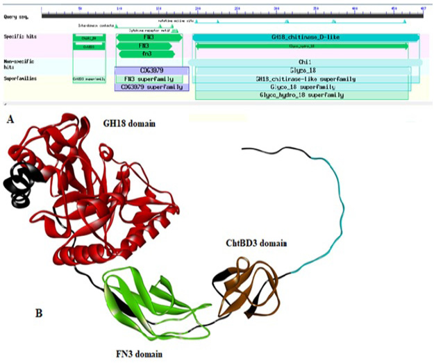
(A) Domain architecture of chitinase of Paenibacillus sp. Y412MC10 by using DbCAN. The locations of the following domains are indicated as: ChtBD3 (aa: 41-83); Fibronectin type III (FN3; aa: 97-173); GH18 (aa: 194-468). (B) 3D structure of chitinase enzyme showing its all domains in respective color.
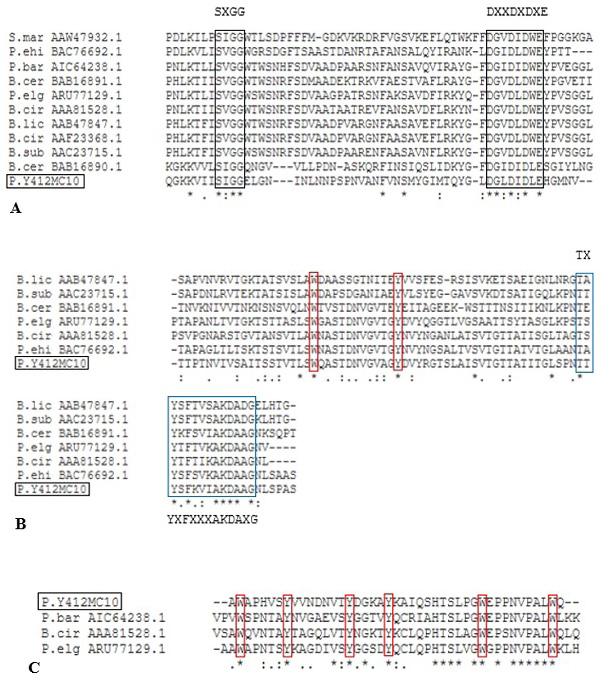
Alignment of putative CatD (A), Fn3D (B) ChBD3 (C) of Paenibacillus sp. Y412MC10 with different characterized bacterial chitinases. Black box indicates the conserved residues in GH 18 catalytic domain. Blue box indicates the conserved residues in Fn3 domain. Red box indicates the conserved aromatic residues W= Tryptophan and Y= Tyrosine, whereas, X may be any amino acid.
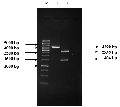
1% Agarose gel electrophoretic analysis of pRSETB/chitinase vector restriction digestion M: 1 kb DNA marker (Fermentas #SM0313); Lane 1: Recombinant plasmid (4299 bp); Lane 2: Double digested plasmid: 2835 bp pRSETB and 1464 bp chitinase gene.
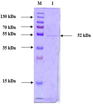
Purified protein SDS-PAGE analysis following anion exchange chromatography. Lane M: Prestained Protein Ladder (PageRuler™ #26616); Lane 1: Purified chitinase.
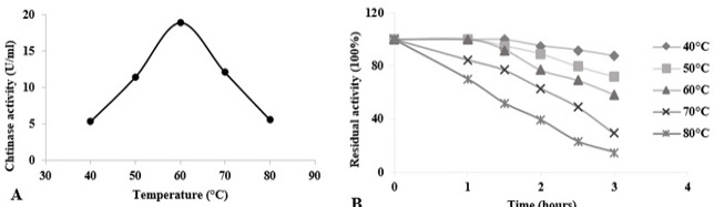
Temperature effects on chitinase activity and stability of Paenibacillus sp. Y412MC10 towards colloidal chitin at pH 5.5. (A) Assessment of optimal temperature for chitinase activity. (B) Evaluation of chitinase thermostability at temperatures from 40 °C to 80 °C. Activity of the non-heated enzyme was defined as 100%.
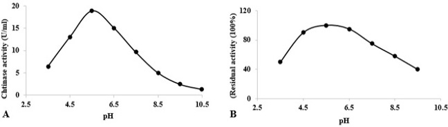
At varying pH values, stability and activity of chitinase towards colloidal chitin. (A) Determination of optimum pH for chitinase activity at 60°C. (B) Stability of chitinase after heating at different pH values for 1 h at 60°C. The activity of the enzyme after incubation at pH 5.5 (in acetate buffer) was defined as 100%.
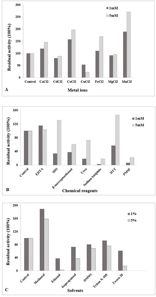
(A) Influence of metal ions on chitinase activity at different concentration (1 mM and 5 mM) of divalent cations. (B) Effect of chemical reagents on chitinase activity at 1 mM and 5 mM concentration. (C) Effect of solvents on chitinase activity at different percentages (1%, 5%). The enzyme was assayed at 60 °C and pH 5.5. Untreated enzyme was used as the control (defined as 100% activity). The data is representative of the mean of three experiments.
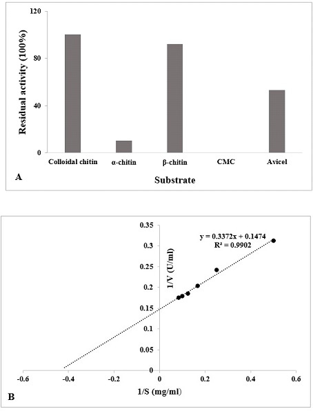
(A) Specificity of different substrate (1%) with the chitinase enzyme was determined (60°C, pH 5.5). The data is represented as % of activity relative to colloidal chitin. (B) Lineweaver-Burk double reciprocal plot of purified chitinase.
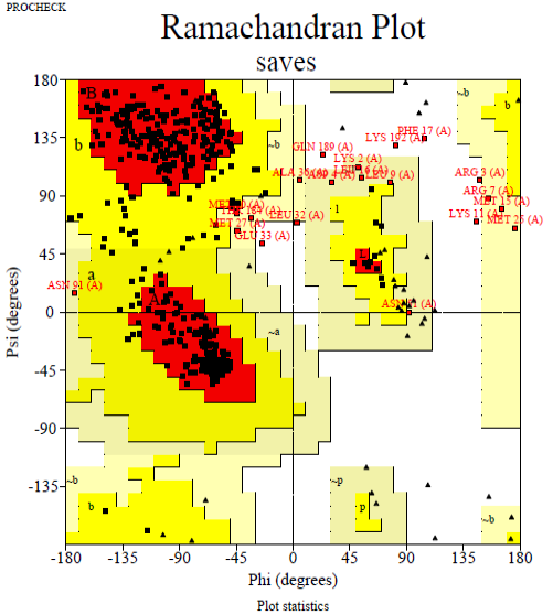
The Ramachandran plot of predicted chitinase indicating total residues in the the most favored areas (red region) and additional additional permitted areas regions (yellow region) are 82.9 and 12.3%, respectively.
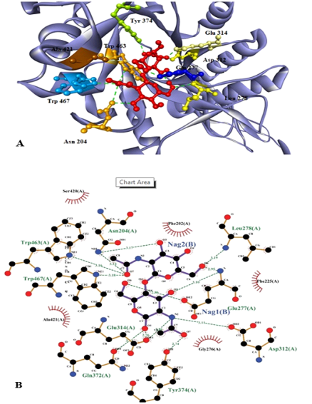
Substrate (NAG)2 docked with chitinase enzyme. Residues are named and the interactions are shown as green dotted lines. (A) 3D structure of the protein with substrate. Red molecules represents 2 molecules of NAG. (B) 2D structure of substrate (NAG)2 docked with chitinase enzyme which shows that active sites involve the interaction of 9 amino acids with 2 molecules of NAG.
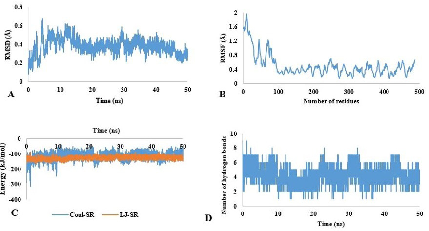
Molecular dynamic simulation of chitinase. (A) RMSD and (B) RMSF fluctuation values for chitinase. (C) Energy values of chitinase for 50 ns. (D) Hydrogen bonds (HB) depiction of chitinase-substrate complex.
References
- Abraham, M.J., Murtola, T., Schulz, R., Páll, S., Smith, J.C., Hess, B., and Lindahl, E.J.S., 2015. Gromacs: High performance molecular simulations through multi-level parallelism from laptops to supercomputers. Software X., 1-2: 19-25. https://doi.org/10.1016/j.softx.2015.06.001[CrossRef]
- Andreini, C., Bertini, I., Cavallaro, G., Holliday, G.L., and Thornton, J.M., 2008. Metal ions in biological catalysis from enzyme databases to general principles. J. Biol. Inorg. Chem., 13: 1205-1218. https://doi.org/10.1007/s00775-008-0404-5[CrossRef]
- Beier, S., and Bertilsson, S., 2013. Bacterial chitin degradation mechanisms and ecophysiological strategies. Front. Microbiol., 4: 149. https://doi.org/10.3389/fmicb.2013.00149[CrossRef]
- Berman, H.M., Battistuz, T., Bhat, T.N., Bluhm, W.F., Bourne, P.E., Burkhardt, K., and Jain, S., 2002. The protein data bank. Acta Crystallogr. D Biol. Crystallogr., 58: 899-907. https://doi.org/10.1107/S0907444902003451[CrossRef]
- Bouacem, K., Laribi-Habchi, H., Mechri, S., Hacene, H., Jaouadi, B., and Bouanane-Darenfed, A., 2018. Biochemical characterization of a novel thermostable chitinase from Hydrogenophilus hirschii strain KB-DZ44. Int. J. Biol. Macromol., 106: 338-350. https://doi.org/10.1016/j.ijbiomac.2017.08.026[CrossRef]
- Bradford, M.M., 1976. A rapid and sensitive method for the quantitation of microgram quantities of protein utilizing the principle of protein dye binding. Anal. Biochem., 72: 248-254. https://doi.org/10.1016/0003-2697(76)90527-3[CrossRef]
- Chaplin, M.F., and Bucke, C., 1990. Enzyme technology. Cambridge University Press. pp. 12.
- Colovos, C., and Yeates, T.O., 1993. Verification of protein structures: patterns of nonbonded atomic interactions. Protein Sci., 2: 1511-1519. https://doi.org/10.1002/pro.5560020916[CrossRef]
- Deng, J.J., Shi, D., Mao, H.H., Li, Z.W., Liang, S., Ke, Y., Luo, X.C., 2019. Heterologous expression and characterization of an antifungal chitinase (Chit46) from Trichoderma harzianum GIM 3.442 and its application in colloidal chitin conversion. Int. J. Biol. Macromol., 134: 113-121. https://doi.org/10.1016/j.ijbiomac.2019.04.177[CrossRef]
- Doan, C.T., Tran, T.N., and Wang, S.L., 2021. Production of thermophilic chitinase by Paenibacillus sp. TKU052 by bioprocessing of chitinous fishery wastes and its application in N-acetyl-D-glucosamine production. Polymers, 13: 3048. https://doi.org/10.3390/polym13183048[CrossRef]
- Eisenberg, D., Lüthy, R., and Bowie, J.U., 1997. VERIFY3D: assessment of protein models with three-dimensional profiles. Methods Enzymol., 277: 396-404. https://doi.org/10.1016/S0076-6879(97)77022-8[CrossRef]
- Fu, X., Yan, Q., Yang, S., Yang, X., Guo, Y., and Jiang, Z., 2014. An acidic, thermostable exochitinase with β-N-acetylglucosaminidase activity from Paenibacillus barengoltzii converting chitin to N-acetylglucosamine. Biotechnol. Biofuels, 7: 174. https://doi.org/10.1186/s13068-014-0174-y[CrossRef]
- Gangwar, M., Singh, V., Pandey, A.K., Tripathi, C.K.M., and Mishra B.N., 2016. Purification and characterization of chitinase from Streptomyces violascens NRRL B2700. Indian J. exp. Biol., 54: 64-71.
- Gao, L., Sun, J., Secundo, F., Gao, X., Xue, C., and Mao, X., 2018. Cloning, characterization and substrate degradation mode of a novel chitinase from Streptomyces albolongus ATCC 27414. Fd. Chem., 261: 329-336. https://doi.org/10.1016/j.foodchem.2018.04.068[CrossRef]
- Gasteiger, E., Hoogland, C., Gattiker, A., Duvaud, S., Wilkins, M.R., Appel, R.D., and Bairoch, A., 2005. Protein identification and analysis tools on the ExPASy server. In: The proteomics protocols handbook (ed. M.J. Walker). Humana Press. https://doi.org/10.1385/1-59259-890-0:571[CrossRef]
- Gopalakrishnan, K., Sowmiya, G., Sheik, S., and Sekar, K., 2007. Ramachandran plot on the web (2.0). Protein Pept. Lett., 14: 669-671. https://doi.org/10.2174/092986607781483912[CrossRef]
- Hamid, R., Khan, M.A., Ahmad, M., Ahmad, M.M., Abdin, M.Z., Musarrat, J., and Javed, S., 2013. Chitinases: An update. J. Pharm. Biol. All. Sci., 5: 21-29. https://doi.org/10.4103/0975-7406.106559[CrossRef]
- Higgins, D.G., and Sharp, P.M., 1988. Clustal: A package for performing multiple sequence alignment on a microcomputer. Gene, 73: 237-44. https://doi.org/10.1016/0378-1119(88)90330-7[CrossRef]
- Jaouadi, N.Z., Rekik, H., Badis, A., Trabelsi, S., Belhoul, M., Yahiaoui, A.B., and Jaouadi, B., 2013. Biochemical and molecular characterization of a serine keratinase from Brevibacillus brevis US575 with promising keratin-biodegradation and hide-dehairing activities. PLoS One, 8: e76722
- Karthik, N., Binod, P., and Pandey, A., 2015. Purification and characterisation of an acidic and antifungal chitinase produced by a Streptomyces sp. Bioresour. Technol., 188: 195-201. https://doi.org/10.1016/j.biortech.2015.03.006[CrossRef]
- Kikkawa, Y., Fukuda, M., Kashiwada, A., Matsuda, K., Kanesato, M., Wada, M., Imanaka, T., and Tanaka, T., 2011. Binding ability of chitinase onto cellulose: An atomic force microscopy study. Polym. J., 43: 742-744. https://doi.org/10.1038/pj.2011.60[CrossRef]
- Kim, Y.H., Park, S.K., Hur, J.Y. and Kim, Y.C., 2017. Purification and characterization of a major extracellular chitinase from a biocontrol bacterium, Paenibacillus elgii HOA73. Pl. Pathol. J., 33: 318-328.
- Komi, D.E., and Hamblin, M.R., 2016. Chitin and chitosan: Production and application of versatile biomedical nanomaterials. Int. J. Adv. Res., 4: 411-427.
- Laemmli, U.K., 1970. Cleavage of structural proteins during the assembly of the head of bacteriophage T4. Nature, 227: 680-685. https://doi.org/10.1038/227680a0[CrossRef]
- Le, B., and Yang, S.H., 2019. Microbial chitinases: Properties, current state and biotechnological applications. World J. Micro. Biotech., 35: 144. https://doi.org/10.1007/s11274-019-2721-y[CrossRef]
- Liaqat, F., and Eltem, R., 2018. Chitooligosaccharides and their biological activities: A comprehensive review. Carbo Poly., 184: 243-259. https://doi.org/10.1016/j.carbpol.2017.12.067[CrossRef]
- Liu, C., Shen, N., Wu, J., Jiang, M., Shi, S., Wang, J., Wei, Y., and Yang, L., 2020a. Cloning, expression and characterization of a chitinase from Paenibacillus chitinolyticus strain UMBR 0002. PeerJ, 8: e8964. https://doi.org/10.7717/peerj.8964[CrossRef]
- Liu, Y., Grimm, M., Dai, W.T., Hou, M.C., Xiao Z.X., and Cao Y., 2020b. CB-Dock: A web server for cavity detection-guided protein–ligand blind docking. Acta Pharm. Sin., 41: 138-144. https://doi.org/10.1038/s41401-019-0228-6[CrossRef]
- Lombard, V., Golaconda, R.H., Drula, E., Coutinho, P.M., and Henrissat, B., 2014. The carbohydrate-active enzymes database (CAZy) in 2013. Nucl. Acids Res., 42: 490-495. https://doi.org/10.1093/nar/gkt1178[CrossRef]
- Loni, P.P., Patil, J.U., Phugare, S.S., and Bajekal, S.S., 2014. Purification and characterization of alkaline chitinase from Paenibacillus pasadenensis NCIM 5434. J. Basic Microbiol., 54: 1080-1089. https://doi.org/10.1002/jobm.201300533[CrossRef]
- Martínez, J.P., Falomir, M.P., and Gozalbo, D., 2014. Chitin: A structural biopolysaccharide with multiple applications. eLS. https://doi.org/10.1002/9780470015902.a0000694.pub3[CrossRef]
- Meng, X.Y., Zhang, H.X., Mezei, M., andCui, M., 2011. Molecular docking: A powerful approach for structure based drug discovery. Curr. Comp. Aided Drug Des., 7: 146-157. https://doi.org/10.2174/157340911795677602[CrossRef]
- Miller, G.L., 1959. Use of dinitrosalicylic acid reagent for determination of reducing sugar. Anal. Chem., 31: 426-428. https://doi.org/10.1021/ac60147a030[CrossRef]
- Mirdita, M., Schütze, K., Moriwaki, Y., Heo, L., Ovchinnikov, S., and Steinegger, M., 2021. ColabFold-Making protein folding accessible to all. https://doi.org/10.1101/2021.08.15.456425[CrossRef]
- Moore, K.G., Price, M.S., Boston, R.S., Weissinger A.K., and Payne, G., 2004. A chitinase from tex 6 maize kernels inhibits growth of Aspergillus flavus. Phytopathology, 94: 82-87. https://doi.org/10.1094/PHYTO.2004.94.1.82[CrossRef]
- Murthy, N.K.S., and Bleakley, B.H., 2017. Simplified method of preparing colloidal chitin used for screening of chitinase-producing microorganisms. Internet J. Microbiol., 10: Corpus ID 13823697.
- Rahman, M.A., Choi, Y.H., Pradeep, G.C., and Yoo, J.C., 2014. An ammonium sulfate sensitive chitinase from Streptomyces sp. CS501. Arch. Pharm. Res., 37: 1522-1529. https://doi.org/10.1007/s12272-014-0509-z[CrossRef]
- Rameshthangam, P., Solairaj, D., Arunachalam, G., and Ramasamy, P., 2018. Chitin and Chitinases: biomedical and environmental applications of chitin and its derivatives. J. Enzymes, 1: 20-43. https://doi.org/10.14302/issn.2690-4829.jen-18-2043[CrossRef]
- Seo, D.J., Lee, Y.S., Kim, K.Y., and Jung, W.J., 2016. Antifungal activity of chitinase obtained from Paenibacillus ehimensis MA2012 against conidial of Collectotrichum gloeosporioides in vitro. Microb. Pathog., 96: 10-14. https://doi.org/10.1016/j.micpath.2016.04.016[CrossRef]
- Stoykov, M.Y., Pavlov, A.I., and Krastanov, A.I., 2015. Chitinase biotechnology: production, purification, and application. Eng. Life Sci., 15: 30-38. https://doi.org/10.1002/elsc.201400173[CrossRef]
- Tanaka, T., Fukui, T., and Imanaka, T., 2001. Different cleavage specificities of the dual catalytic domains in chitinase from the hyperthermophilic archaeon Thermococcus kodakaraensis KOD1. J. biol. Chem., 276: 35629-35635. https://doi.org/10.1074/jbc.M105919200[CrossRef]
- Trott, O., and Olson, A.J., 2010. Auto Dock Vina: improving the speed and accuracy of docking with a new scoring function, efficient optimization, and multithreading. J. Comput. Chem., 31: 455-461. https://doi.org/10.1002/jcc.21334[CrossRef]
- Tuveng, T.R., Hagen, L.H., Mekasha, S., Frank, J., Arntzen, M.O., Vaaje-Kolstad, G., and Eijsink, V.G.H., 2017. Genomic, proteomic and biochemical analysis of the chitinolytic machinery of Serratia marcescens BJL200. Biochim. biophys. Acta Proteins Proteom., 1865: 414-421. https://doi.org/10.1016/j.bbapap.2017.01.007[CrossRef]
- Ueda, J., and Kurosawa, N., 2015. Characterization of an extracellular thermophilic chitinase from Paenibacillus thermoaerophilus strain TC22-2b isolated from compost. World J. Microbiol. Biotechnol., 31: 135-143. https://doi.org/10.1007/s11274-014-1754-5[CrossRef]
- Vaaje-Kolstad, G., Horn, S.J., Sorlie, M., and Eijsink, V.G.H., 2013. The chitinolytic machinery of Serratia marcescens - A model system for enzymatic degradation of recalcitrant polysaccharides. FEBS J., 280: 3028-3049. https://doi.org/10.1111/febs.12181[CrossRef]
- Wang, D., Li, A., Han, H., Liu, T., and Yang, Q., 2018. A potent chitinase from Bacillus subtilis for the efficient bioconversion of chitin-containing wastes. Int. J. Biol. Macromol., 116: 863-868. https://doi.org/10.1016/j.ijbiomac.2018.05.122[CrossRef]
- Waterhouse, A., Bertoni, M., Bienert, S., Studer, G., Tauriello, G., Gumienny, R., Heer, F.T., de Beer, T.A.P., Rempfer, C., Bordoli, L., Lepore, R., and Schwede, T., 2018. Swiss-model: Homology modelling of protein structures and complexes. Nucl. Acids Res., 46: W296-W303. https://doi.org/10.1093/nar/gky427[CrossRef]
- Yan, Q., and Fong, S.S. 2018. Cloning and characterization of a chitinase from Thermobifida fusca reveals Tfu_0580 as a thermostable and acidic endochitinase. Biotechnol. Rep., 19: e00274. https://doi.org/10.1016/j.btre.2018.e00274[CrossRef]
- Yang, S., Fu, X., Yan, Q., Guo, Y., Liu, Z. and Jiang, Z., 2016. Cloning, expression, purification and application of a novel chitinase from a thermophilic marine bacterium Paenibacillus barengoltzii. Fd. Chem., 192: 1041-1048.
- Yin, Y., Mao, X., Yang, J.C., Chen, X., Mao, F., and Xu, Y., 2012. dbCAN: A web resource for automated carbohydrate-active enzyme annotation, Nucl. Acids Res., 40(Web Server issue): W445-451. https://doi.org/10.1093/nar/gks479[CrossRef]
- Younas, A., Saleem, M., Tariq, H., Arooj, B., Akhthar, M.S., and Tahira, R., 2016. Purification and biochemical characterization of a pathogenesis related endochitinase Arachis hypogaea. Int. J. Agric. Biol., 18: 780-788. https://doi.org/10.17957/IJAB/15.0167[CrossRef]
- Yu, W., He, X., Vanommeslaeghe, K., and MacKerell Jr, A.D., 2012. Extension of the CHARMM general force field to sulfonyl-containing compounds and its utility in biomolecular simulations. J. Comput. Chem., 33: 2451-2468. https://doi.org/10.1002/jcc.23067[CrossRef]
- Zhang, A., He, Y., Wei, G., Zhou, J., Dong, W., Chen, K., and Ouyang, P., 2018. Molecular characterization of a novel chitinase CmChi1 from Chitinolyticbacter meiyuanensis SYBC-H1 and its use in N-acetyl-D-glucosamine production. Biotechnol. Biof., 11: 179. https://doi.org/10.1186/s13068-018-1169-x[CrossRef]
- Zhou, J., Chen, J., Xu, N., Zhang, A., Chen, K., Xin, F., Zhang, W., Ma, Z., Fang, Y., Jiang, M., and Dong, W., 2019. The broad-specificity chitinases: Their origin, characterization, and potential application. Appl. Microbiol. Biotechnol., 103: 3289-3295. https://doi.org/10.1007/s00253-019-09718-x[CrossRef]
Services
Article usage statistics
Loading…