
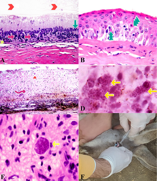
Eye, change in Traumatic Brain Injury. A: Mild degeneration of Retina, including loss of cone and rod photoreceptor processes, (yellow arrowheads), unicellular necrosis (single-cell) in the outer part of nuclear layer photoreceptor cells, (Pink Arrows), Disorganization and hypo cellularity of the outer and inner parts of the nuclear layers, (Green Arrow) and narrowing and absence of the plexiform layers. (redhead arrow). (H & E, x800). B: Cornea ,Necrosis. Hypereosinophilic and shrunken necrotic cells, (Green Arrow) (H & E, x800). C: overlying necrosis of retina, (Pink Arrow) and presence of a large Toxoplasma gondii tissue pseudocyst on the surface. (redhead arrow) (H & E, x600). D, E: Toxoplasma gondii cyst-containing discrete granule-type bradyzoites (yellow arrows) (H & E, x800). F: Intraocular injection in another patient kangaroo.