Figure 2:
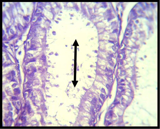
A histopathological slice of the first group’s testes at 4 weeks reveals severe vacuolations of the seminiferous tubular epaithelium with the lack of spermatogenic cells (black arrow) (H & E stain X 40).
A histopathological slice of the first group’s testes at 4 weeks reveals severe vacuolations of the seminiferous tubular epaithelium with the lack of spermatogenic cells (black arrow) (H & E stain X 40).