
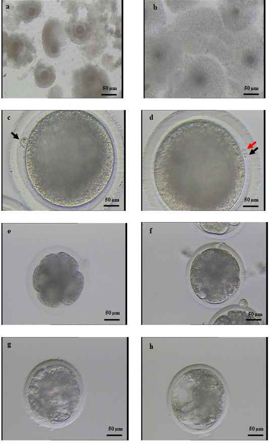
Figure 1:
Representative image of embryonic development from bovine oocytes using cysteine supplementation during IVM. (a) Immature cumulus-oocyte complexes; (b) Cumulus expansion at 22 h after culturing in IVM medium; (c) Matured oocyte with a polar corpuscle (black arrow); (d) Zygote with two polar bodies (red and black arrows); (e) 8-cells embryo at 72 h after IVC; (f) 16-cells embryo at 96 h after IVC; (g) Early blastocyst at 156 h after IVC; (h) blastocyst at 168 h after IVC.