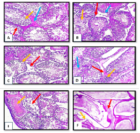
Figure 1:
Histopathological section of testes and epididymis tissue from tetrodotoxin treated group (0.5 µg/kg.bw) of male albino rats (A) sever degenerated changes of spermatogonia cells (red arrow) with irregular appearance of spermatid (yellow arrow) with presence of edematous substance between degenerated seminiferous tubules. (B) obvious disruption of seminiferous tubules (red arrow) with necrotic finding of some tubules (yellow arrow) with absence of leydig cells (blue arrow). (C) complete loss of leydig cells (red arrow) with evidence of hypospematogensis (yellow arrow). (D) intertubler edema (red arrow) with mild mononuclear cells infiltration (yellow arrow) with necrotic of immature spermatid (blue arrow). complete necrosis of spermatogonial cells (red arrow) with obvious subcapsulear vascular congestion and dilation (yellow arrow). (E) complete necrosis of spermatogonial cells (red arrow) with obvious subcapsulear vascular congestion and dilation (yellow arrow). H&E, x10. (F) marked irregularity of epididymal tubules (red arrow) with obvious atrophy of some tubules (yellow arrow). H&E, x40