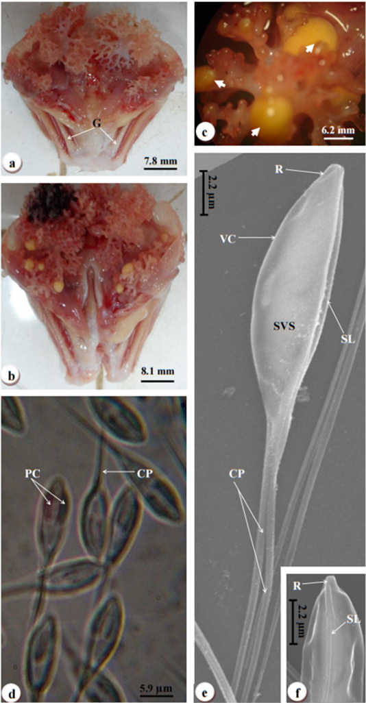
Digital camera photographs illustrating various aspects of the ABO infection. (a) Uninfected ABO demonstrating organ support by the gill arches (G). (b) Infected ABO displaying multiple cysts (thick arrowheads) at the bulb’s tip. (c) Infected ABO showcasing large yellowish cysts (white arrowheads) occupying the tip of the bulbs and the stems of the ABO. (d) Photomicrograph of fresh myxospores of Henneguya sp., revealing the spore body (Sp), two pyriform polar capsules (PC), and the caudal processes (CP). (e) Scanning electron microscope photomicrographs of the myxospores. (f) Close-up highlighting the smooth valvular surface (SVS) of the myxospore, the sutural line (SL), and the caudal processes (CP) extending from the valves, as well as the rostrum (R) extending from its anterior end. (g) Profile view of the anterior end of the myxospore, clearly showing the sutural line (SL) and the rostrum (R).
