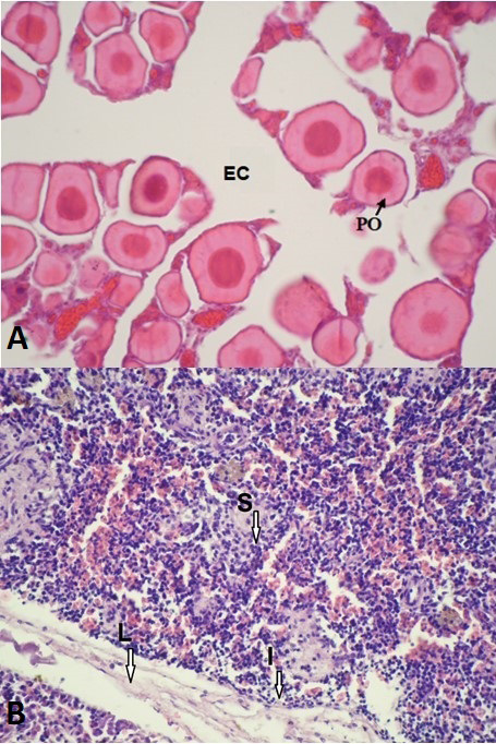
Fig. 1.
Histological structure of ovary (A) and tests (B) of Clarias gariepinus. S, spermatids; L, Lumen; I, Interstitial tissue; PO, Primary oocytes; EC, Endoovarian canal) Stain: H & E. Magnification: A, 10X; B 40X scale bar: A, 50 µm; B, 100 µm.