
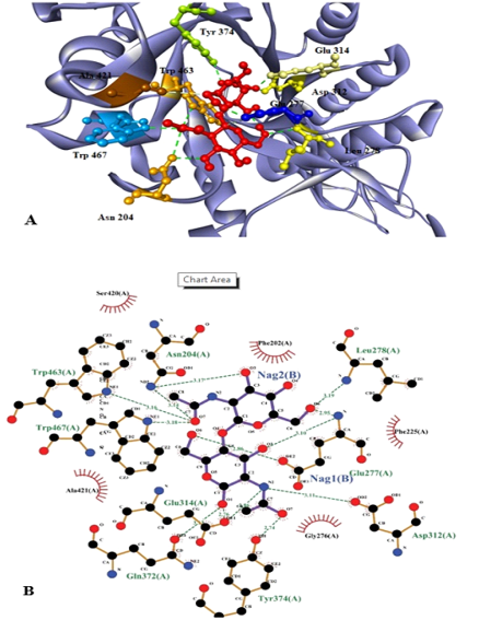
Fig. 10.
Substrate (NAG)2 docked with chitinase enzyme. Residues are named and the interactions are shown as green dotted lines. (A) 3D structure of the protein with substrate. Red molecules represents 2 molecules of NAG. (B) 2D structure of substrate (NAG)2 docked with chitinase enzyme which shows that active sites involve the interaction of 9 amino acids with 2 molecules of NAG.