
Figure 8:
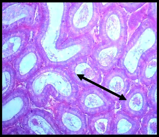
Histopathological slice of the second group’s testes at 4 weeks exhibits hyperplasia of clear cells that are strongly vacuolated, homogeneous material, and cellular debris (black arrow) in the lumen (H & E stain X 40).
Histopathological slice of the second group’s testes at 4 weeks exhibits hyperplasia of clear cells that are strongly vacuolated, homogeneous material, and cellular debris (black arrow) in the lumen (H & E stain X 40).