
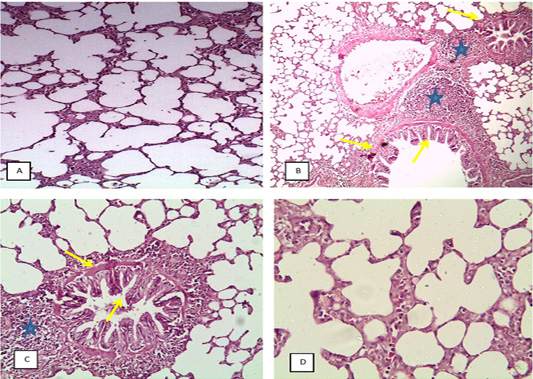
Figure 7:
(H&E X4), A: lung of CG showing alveolar emphysema, B: lung of TG showing bronchitis and bronchiolitis, peribronchial edema, hyperplasia of epithelial lining (yellow arrow) and sever peribronchial infiltration of chronic inflammatory cells (star) and alveolar emphysema, C: lung of DAG showing catarrhal bronchiolitis, hyperplasia of epithelial lining, edema (yellow arrow) with massive infiltration of chronic inflammatory cells, (star) and alveolar emphysema, GD: lung of BG beginning to return to normal showing alveolar emphysema.