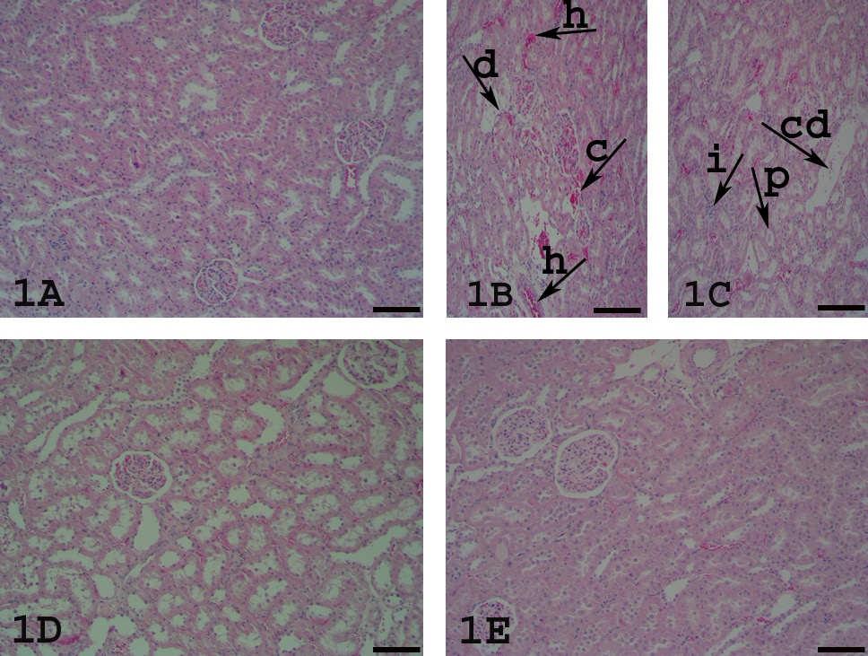
Fig. 1.
Histopathological appearance of kidney tissues of all groups, HE staining, scale bar= 150 µm A, Control group: normal kidney morphology. B-C, CP group: hyperemia, epithelial degeneration, swelling, cystic dilatation, proteinaceous material in tubuli, congested and hypercellular glomeruli, inflammatory cells infiltration in cortical and medullary area. Arrows pointing events; h: hyperemia, c: congestion, d: degeneration, i: inflammation, p: proteinaceous material, cd: cystic dilatation. 1D, CP+EA50 group: significant decrease in pathological lesions. 1E, CP+EA75 group: more protective effect and normal architecture of kidney.