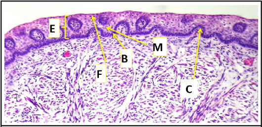
Figure 7:
Cross histological section of body of the tongue sheep fetuses at (70-75) day showing: epithelial layer; a basal layer of cuboidal cells (B) which dark stain in color, middle layer of squamous cells (M) or hexagonal cells lighter in color with irregular or rounded centrally nuclei light in color and finally, superficial layer including flattened cells (F) may be keratin or non-keratin on dorsum and invagination of connective tissue core (C) (H & E stain, 10X).
