
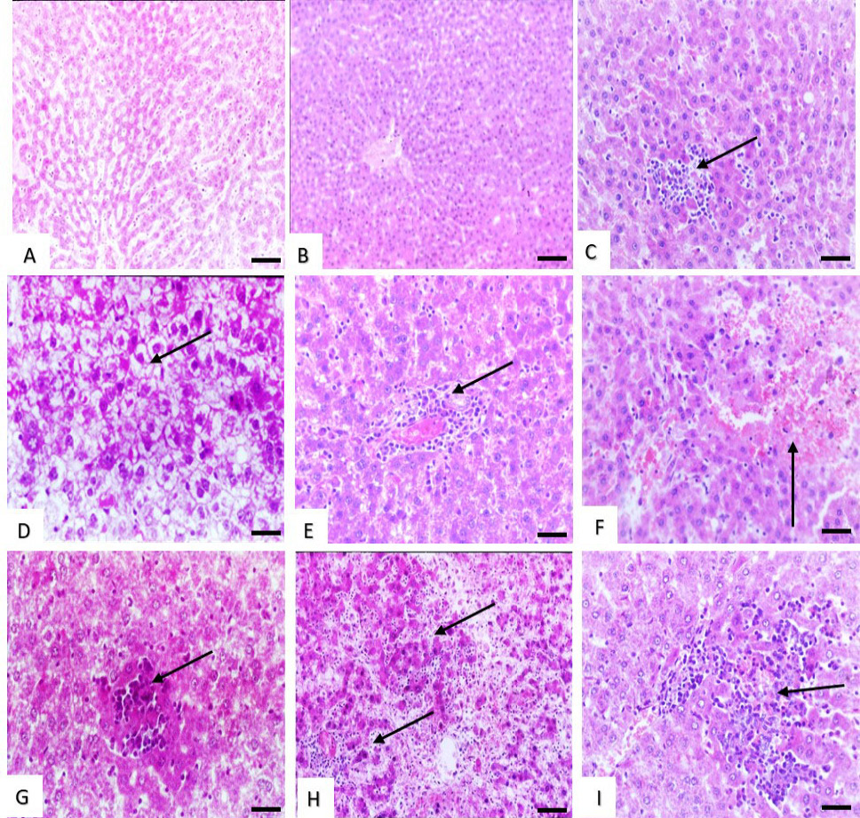
Histological structure of rat liver (A and B) Effect of low dose of malathion and metalaxyl showing, slight congestion and dilation of the hepatic sinusoids. (C) Effect of low dose of cymoxanil showing slight sinusoidal cell activation with pregranuloma formation (arrow). (D) Effect of medium dose of malathion showing marked cytosolic hydrops with vacuolated cytoplasm of the hepatocytes (arrow). (E) Effect of medium dose of metalaxyl showing perivascular focal collection of mononuclear cells (arrow). (F) Effect of medium dose of cymoxanil showing haemorrhages in the hepatic parenchyma (arrow). (G and H) Effect of high dose of malathion, metalaxyl and cymoxanil showing hepatocellular necrosis (arrows). Stain: H and E. Magnification bar: 50 µm.