
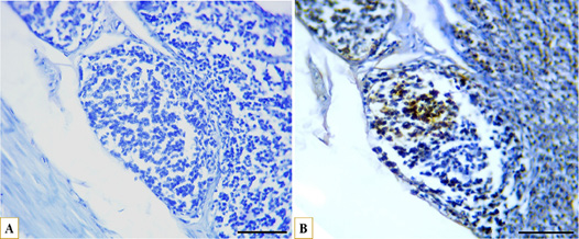
Figure 11:
Microphotographs showing immunohistochemical expression of CD40 in B lymphocytes in the middle caecal portion and distribution of B lymphocytes (A) control with negative CD40 expression, (B) strong positive expression of CD40 (brown) localized to the cell membrane of B lymphocytes (blue), IHC, scale bar = 50 µm.