
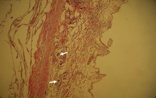
Figure 3:
A section of the intestine infected with nematodes shows the complete destruction of all cellular layers. Hypertrophy of villous crypt is observed whereas villi are completely washed out. Fibrosis of mucosa and sub-mucosa is also obvious. Section of nematode larvae (Ascaridia sp.) observed in this section. (Arrow) × 100.