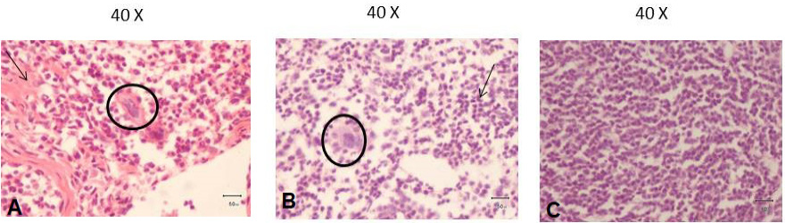
Fig. 4.
Representative images of histopathology of the RB51 and B. abortus ΔpurD mutant in mice. Six out of eight mice from RB51 mice group spleen showed atrophy of follicular area and dead tissue mass is also seen in the (arrow) with few giant cells are also present (circle) (A). Six mice out of eight from B. abortus ΔpurD mice group spleen showed giant phagocytic cells with phagocytized bacteria were seen (circle) and higher infiltration of lymphocytes (arrow) (B). Control mice group spleen showed no tissue changes and almost normal tissue parenchyma was seen (C).