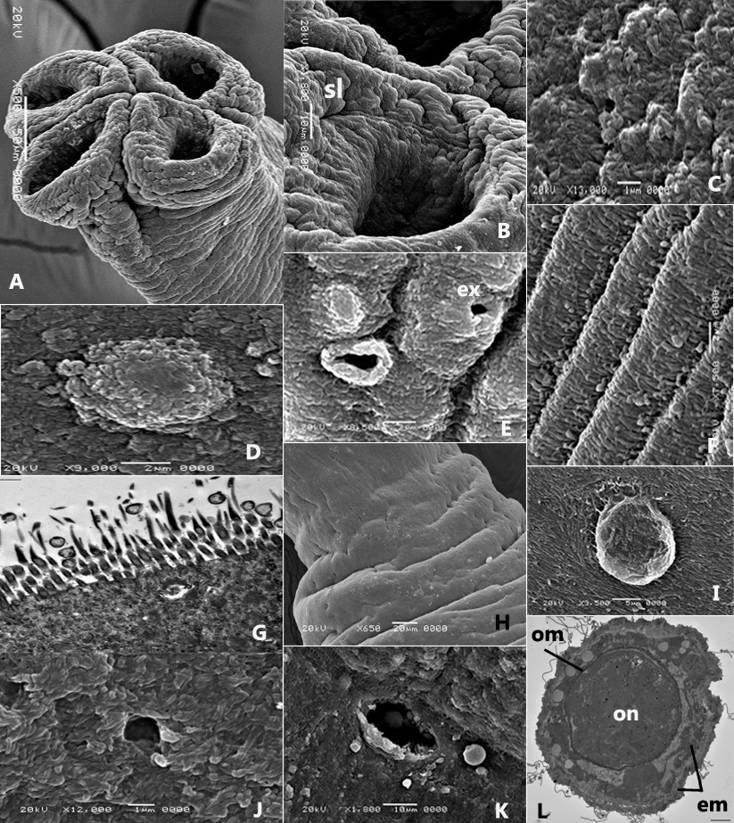
(A-F, H-K) Scanning and, (G, L) Transmission electron micrographs of Ophiotaenia tessellata sp. n. from Natrix tessellata, Egypt. (A) Scolex with 4 suckers showing wrinkles and folds; (B) an enlarged sucker showing the transverse slit-like structure (sl); (C) acicular filitriches covering the proliferative zone; (D) a tumulus on the proliferative zone; (E) Burst of the tumulus on the proliferative zone forming large pore. Note also the excretory pore (ex); (F) acicular filitriches covering immature proglottis; (G) acicular filitriches and few gladiate spinitriches covering mature proglottis; (H) excretory pores scattered on mature proglottis; (I) a tumulus on mature proglottis; (J) acicular filitriches covering gravid proglottis; (K) one uterine pore; (L) an egg in a stage of development showing the oncosphere (on), oncospheral membrane (om) and bi-layered embryophore (em). Scale-bars: A= 50μm; B, K= 10μm; C, J= 1μm; D-F, L= 2μm; G= 500nm; H= 20μm; I= 5μm.