
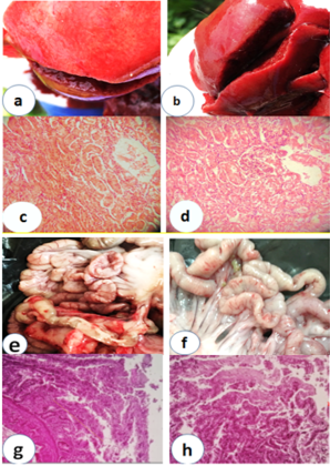
Gross and histological images of the liver and intestine of chickens reared under heat stress. a. Liver showed swelling and multiple spots of congestive pales to whitish areas. b. Liver showed marked congestion, edematous with enlarged rounded edge. C. Liver tissue with cloudy swelling of liver cells with dilatation of sinusoidal portal vein. d. Liver tissue showed dilated liver parenchyma, lymphocytic cuffing, and disorganization of the hepatic cord. e. Small intestine shows congestive blood vessels with petechial hemorrhages. f. Small intestine appeared congestive of blood vessel dilatation and empty lumen of the intestine. g. Small intestine (Duodenum) appears hyperplasia villi, proliferation of epithelial cells with, destructive some intestinal glands h. Small intestine (duodenum) with necrosis and destructive of the cell wall and proliferation of the villi with intestinal catarrhal discharge.