
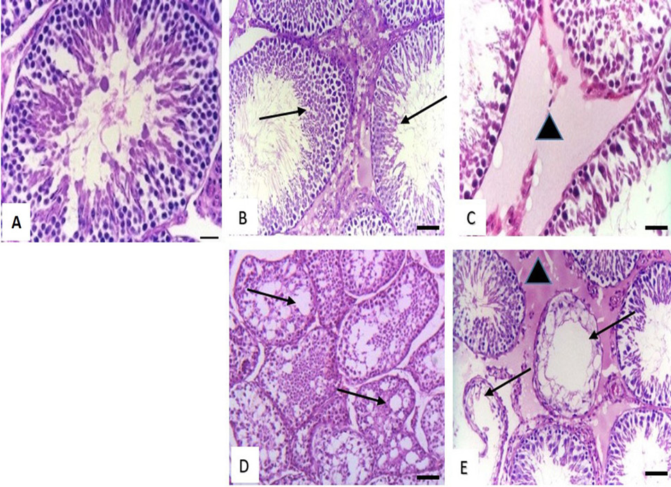
Fig. 5.
Histological structure of rat testis. (A) Effect of low dose of malathion, (low, medium, high) dose of metalaxyl and low dose of cymoxanil showing normal structure. (B) Effect of medium dose of malathion showing decrease in the number of spermatogenic cells in the seminiferous tubules (arrows). (C) Effect of medium dose of cymoxanil showing interstitial oedema (triangle). (D) Effect of high dose of malathion showing necrosis of spermatogenic layers and marked vacuolation in the cytoplasm of sertoli cells (arrows). (E) Effect of high dose of cymoxanil showing, massive necrosis in the lining epithelium of seminiferous tubules (arrows) with marked interstitial oedema (triangle). Stain: H and E. Magnification bar: 50 µm.