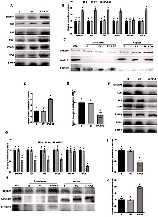
Activation of SREBP1 pathway by RPL8 in YMECs. In A-B, the expression of RPL8, ACC, FAS, SCD1, SREBP1, and PPARγ was evaluated in YMECs subjected to RPL8 overexpression. The nuclear and cytoplasmic localization of SREBP1 was also assessed in cells treated with RPL8 overexpression, as depicted in C-E. The results showed that the expression of these proteins, as well as the nuclear localization of SREBP1, was significantly increased in cells with RPL8 overexpression. Conversely, F-G displays the expression of RPL8, ACC, FAS, SCD1, SREBP1, and PPARγ in YMECs treated with RPL8 silencing. Similarly, H-J demonstrates the nuclear and cytoplasmic localization of SREBP1 in cells treated with RPL8 silencing. The results showed that the expression of these proteins, as well as the nuclear localization of SREBP1, was significantly decreased in cells with RPL8 silencing. In B and G, the expression of proteins in the “B” group was set to “1”. In D and I, the nuclear localization of SREBP1 in the “B” group was set to “1”, while in E and J, the cytoplasmic localization of SREBP1 in the “B” group was also set to “1”. Cells were divided into different groups, including those that were not transfected B, those that were transfected with the empty vector (EV), those that were transfected with the RPL8 overexpression vector (RPL8 GO), those that were treated with whole cell lysate (WCL), those that were transfected with the negative control siRNA (NC), and those that were transfected with the RPL8 siRNA (Si-RPL8). In the bar charts, different superscript letters indicate significant differences (p<0.05), while the same letters represent no significant difference (p>0.05).