Figure 10:
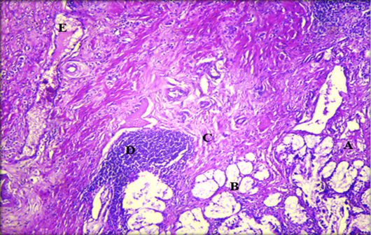
Histopathological section of gallbladder showing A: necrotic tissue; B: destruction of glands; C: fibrosis of mucosa; D: aggregation of lymphocytes cells and E: hemorrhage (H & E stain. X10).
Histopathological section of gallbladder showing A: necrotic tissue; B: destruction of glands; C: fibrosis of mucosa; D: aggregation of lymphocytes cells and E: hemorrhage (H & E stain. X10).