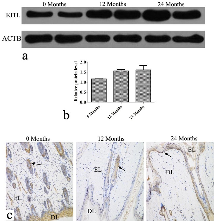
Fig. 4.
KITL protein expression and localization. a, the KITL protein was detected by western blotting. ATCB was used as a loading control. b, relative protein band intensity was determined and quantified by Image J. Data are presented as mean ± SD (n=3). c, immunohistochemical analysis of the KITL protein. EL, epidermal layers; DL, dermal layers. Bar = 50 μm.