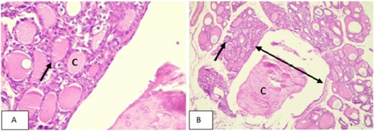
Figure 1:
Photomicrographs of the thyroid of rabbits of negative control group (C1) showed normal architecture of the thyroid gland. It comprised of different-sized thyroid follicles (black arrow) filled with acidophilic homogenous colloid (C), and individual follicle was lined with a simple layer of cubical epithelial cell (Blue arrow). (A)H&E 10X.(B) H&E 40X.
