
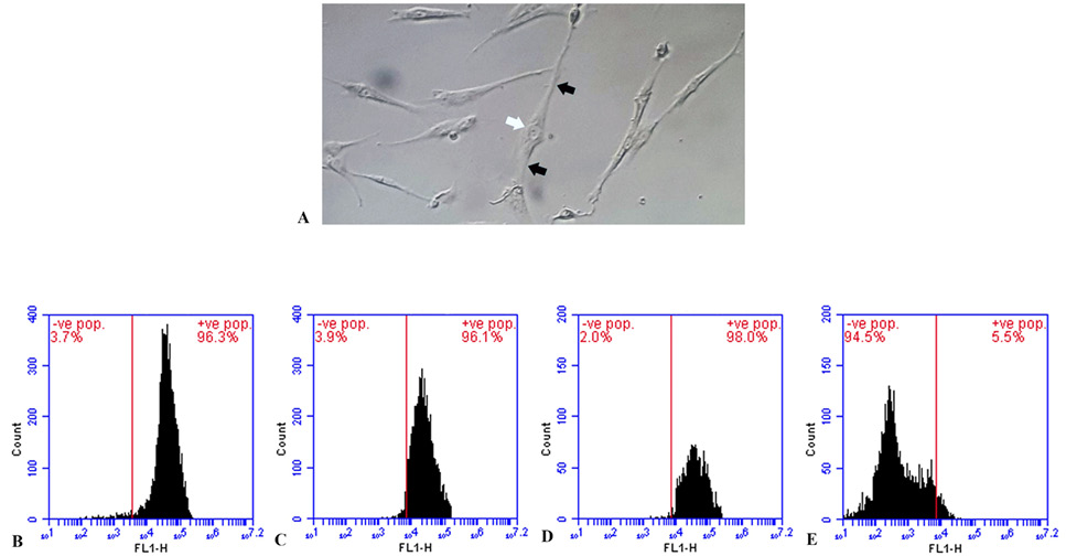
Fig. 1.
The identification of rat Bone marrow mesenchymal stem cell (BMMSCs) by microscopy and flowcytometry. The data shows a microscopic image (A) of BMMSCs, taken after 4 days in culture. The cells appear spindle-shaped, with a central cell body (white arrow) and cytoplasmic processes in opposite directions (black arrows). The data also shows flowcytometric identification of BMMSCs, revealing positive expression of specific surface markers: CD44 (B), CD90 (C), and CD105 (D) and negative expression for hemopoietic and endothelial surface marker CD34 (E).