
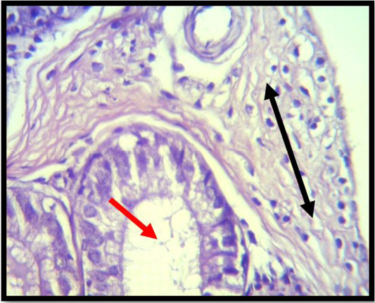
Figure 1:
A histopathological slice of the first group’s epididymes at 4 weeks demonstrates vacuolated cells, poorly differentiated spermatogenic cells (red arrow), and interstitial edema. Interstial tissue with inflammatory cell infiltration (black arrow) (H & E stain X 40).