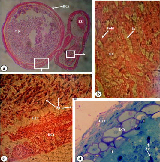
Photomicrographs of different sections from the tip of the ABO bulb, stained with Haematoxylin-Eosin and toluidine blue. (a) Transverse section of an infected bulb tip, stained with Haematoxylin-Eosin, revealing a dense connective tissue (DCt) and elastic cartilage (EC) filled with myxospores (Sp) of Henneguya sp. (x 400). (b) Haematoxylin-Eosin stain of a transverse section in the elastic cartilage (EC) of an uninfected ABO, showing large folds and intervening spaces (Is) (x 600). (c) Haematoxylin-Eosin stain of a transverse section of an infected bulb tip, illustrating the dense connective tissue (DCt), loose connective tissue (LCt), and elastic cartilage (EC). Myxospores were observed within the elastic cartilage (EC) and the LCt (x 600). (d) Toluidine blue stain photomicrograph of an infected bulb tip section of the ABO, demonstrating myxospores (Sp) within the elastic cartilage (EC). Notice the progressive destruction (thick white arrow) of the elastic cartilage (EC) towards the loose connective tissue (LCt) (x 1,000).
