
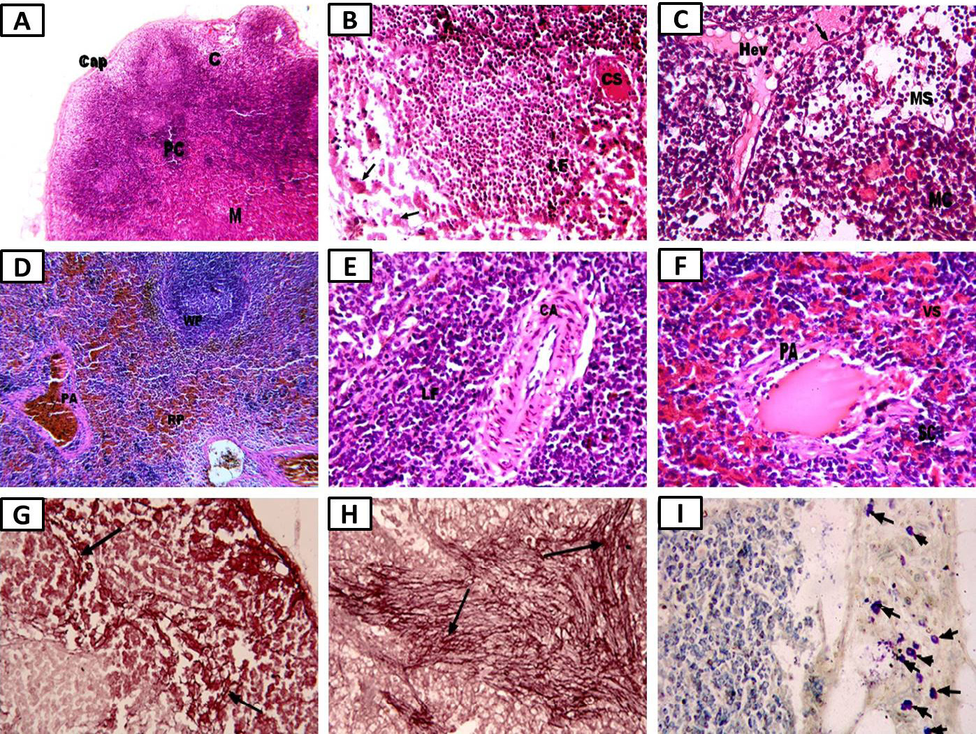
Photomicrographs of the benzene-injected group showing: A, whole lymph node structure (×100); B, cortical changes with some necrotic (ghost) cells (arrow), dilated, congested cortical sinus (CS), and non-active lymphoid follicles (LF) (×400); C, medullary changes with dilated and congested high endothelial venules (HEVs) lined with simple squamous epithelium (arrow) (×400); D, spleen with dilated congested pulp artery (PA) (×100); E, white pulp (WP) with thick dilated central arteriole (CA) in non-active LF (×400); F, red pulp (RP) with congested venous sinuses (VS) and dilated PA (hematoxylin and eosin, ×400); G, densely stained irregular thick elastic fibers (arrow) (×400); H, thick elastic fiber aggregation in the spleen (arrow) (Orcein, ×400); I, increased numbers and larger than normal mast cells (arrows) in the pericapsular connective tissue of the lymph node (toluidine blue, ×400).