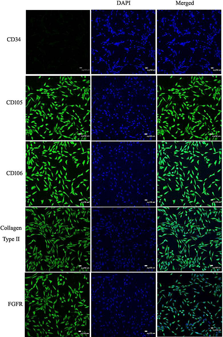
Fig. 3.
Immunofluorescence of CPSC surface markers. Cell nuclei stained with DAPI are shown in the middle panels. The pictures shown above indicate that the CPSCs positively expressed FGFR-3, collagen type II, CD105, and CD106, and negatively expressed CD34 (Scale bar = 50 μm).