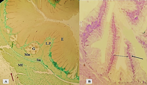
Figure 6:
Photographic section of the thoracic part esophagus in chick turkey, (A): The mucosal folds become, elongated, and unbranched leaving only a narrow lumen, T. serosa (brown arrow) A: Masson’s Trichrome stain A: 100 X, (B) mucus gland in turkey lined by columnar cell with basal nucleus: PAS stain 400 x.
