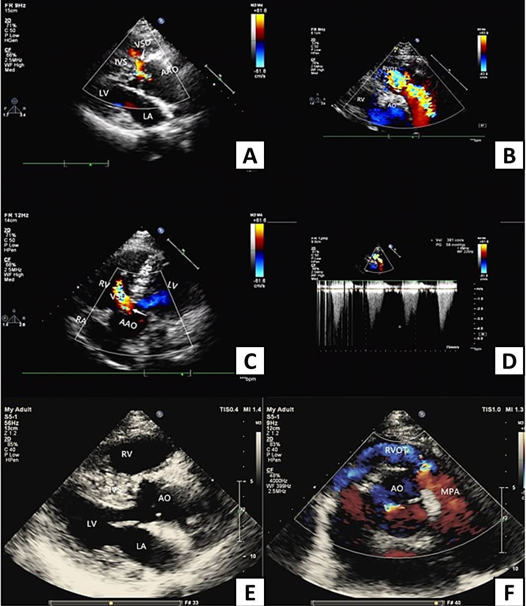
Transthoracic echocardiography before and after corrective surgery (AAO, ascending aorta. AO, aorta; RVOT, right ventricular outflow tract; VSD, ventricular septal defect; IVS, interventricular septum; RA, right atrium; LA, left atrium; RV, right ventricle; LV, left ventricle). A, Parasternal long axis view of left ventricle shows interruption of ventricular septum continuity before surgery, arrow points to left to right shunt of ventricular defect. B, Nonstandard axis view shows obvious acceleration of right ventricular outflow tract blood flow with multicolored mosaic blood flow signals before surgery. C, Apical 5-chamber view shows interruption of ventricular septum continuity before surgery, arrow points to left to right shunt of ventricular defect. D, Color doppler flow diagram of right ventricular outflow tract before surgery, the blood flow of right ventricular outflow tract was obviously accelerated, the peak systolic velocity was about 3.8 m/s, and the pressure difference was 54 mmHg. E, Long axis view of the left ventricle shows RVH, closure of the VSD and RVOT augmentation after corrective surgery. F, The short axis view of the aorta shows the blood flow of right ventricular outflow tract was nearly normal after corrective surgery, no blood flow acceleration was found.