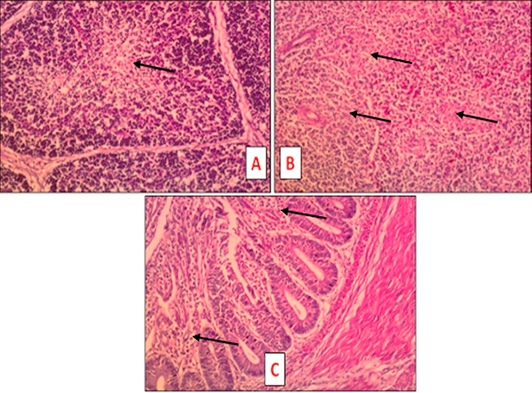
Figure 4:
Tissue sections of S. aureus and /or E. coli infected treated birds at 7 DPT (HandE, X 200) showing the following lesions. A: S. aureus + E. coli infected treated bird: bursa slight depletion of the lymphoid follicle (head of arrow). B: S. aureus + E. coli infected treated bird: spleen slight depletion of the lymphoid follicle (head of arrow). C: S. aureus infected treated bird: intestine light inflammation characterized by lymphocytic infiltration of the mucosa (head of arrow).