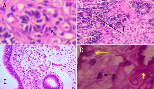
Figure 2:
A: Acute endometritis: The glandular lamina and peri-endometrial glands show mild to moderate infiltration of mononuclear cells (blue arrow). 400X H&E stain. B: Acute endometris: enlargement of endometrial glands with degenerative changes (blue arrow) 100X H&E stain. C: Subacute endometritis: Marked hyperplasia of luminal epithelium (black arrow) with congestion (yellow arrow), edema and infiltration of inflammatory cells in lamina propria with glandular epithelial hyperplasia (blue arrow) (100x H & E). D: Subacute endometritis There was moderate to severe infiltration of mononuclear cells especially plasma cells (black arrow) as well as epithelioid cells in lamina propria around endometrial gland (yellow arrow) 400x H&E stain.
