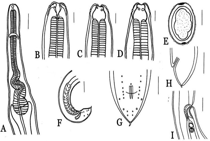
Fig. 1.
Seuratascaris schmackeri sp. nov. from Odorrana schmackeri in China. (A) anterior part of male, laterial view, showing oesphagus, nerve-ring, excretory pore, intestinal caecum. (B) anterior part of male, laterial view, showing interlabial and postlabial groove. (C) anterior part of male, dorsal view, showing dorsal lip with two pipillars. (D) anterior part of female, lateral-ventral view, showing lateral-ventral lip with one papillae and amphid. (E) egg. (F) posterior end of male, lateral view. (G) posterior end of male, ventral view. (H) posterior end of female, lateral view. (I) region of Vulva, lateral view. Scale bars: A= 200μm; B, C, D= 500μm; E= 50μm; F, G, H = 200μm; I = 300μm.