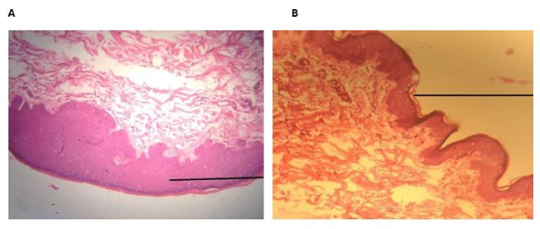
Figure 1:
Histopathological evaluation of wound healed with PTB. The photomicrograph showed maximum leucocytic infiltration. More prominent and regular re-epithelization was seen. Prominent keratinization was seen, thickness of epidermis and dermis was more. H & E; 10X. (B) Histopathological evaluation of wound healed with silk. The silk treated photomicrograph showed less thickness of epithelium as compared to PTB. Thickness of epidermis and dermis was less. No inflammatory cells were seen. H & E; 10X.
