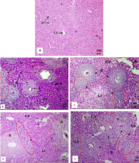
(A-E): Light micrograph of liver section of (A) Normal control mouse showing normal hepatocytes (H) and central vein. (B) S. mansoni infected mouse eight-week post infection showing abnormal hepatocytes (Ab. H), multiple Schistosoma eggs (E) provoke granuloma formation (G) of large diameters containing extended regions of lymphocytic infiltration (L.I). Cytoplasmic vacuolization is clear (C.V). (C) S. mansoni infected mice treated with PZQ drug showing granuloma (G) with high lymphocytic infiltration (L.I), clear cytoplasmic vacuolization (C.V). (D) S. mansoni infected mice treated with FWE showing granuloma (G) with clear lymphocytic infiltration (L.I) and mild cytoplasmic vacuolization (C.V). (E) S. mansoni infected mice treated with PZQ accompanied with FWE showing multiple granuloma (G) with high lymphocytic infiltration (L.I), multiple cytoplasmic vacuolization (C.V). (A-E: H and E X100).