
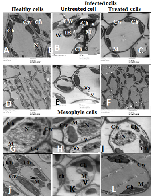
Figure 5:
Ultra-micrograph section in mesophyll tissue of healthy infected and treated fig leaf ChNPs and biomagic showing: (A) Impact cells and (B) Magnified nucleus (N) showing compact nucleus in healthy cell and deformed nucleus in infected cell. CW: cell wall; Ch: chloroplast; V: Virus; Vs: vesicles.