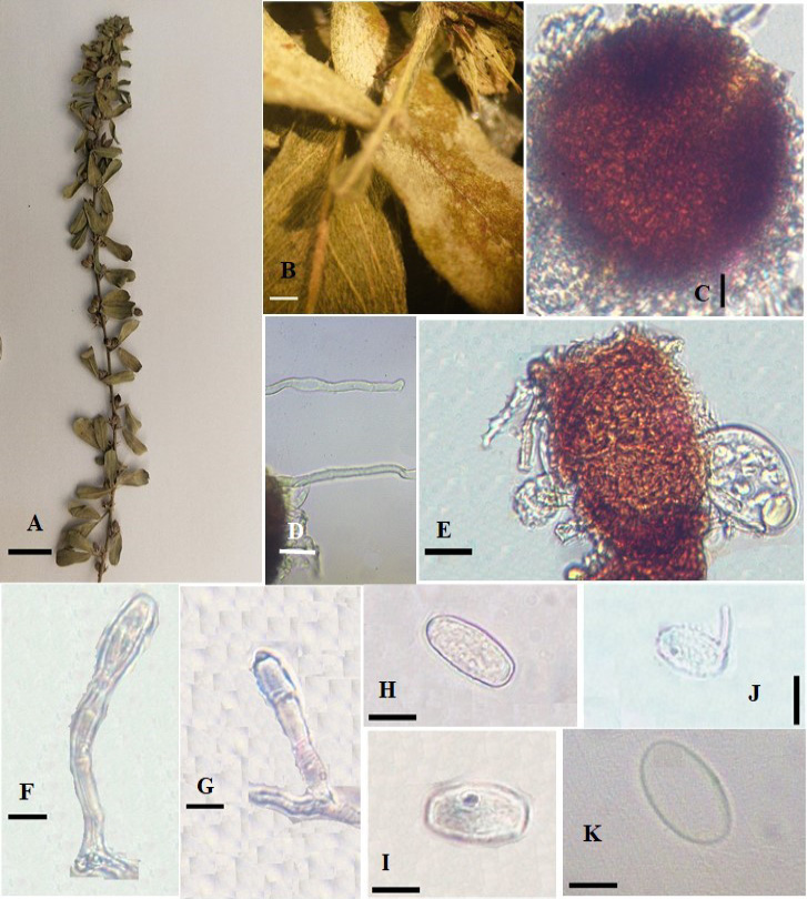
Figure 1:
Erysiphe diffusa on Lespedeza tomentosa A: Infected leaves of Lespedeza tomentosa (AFE022,) B: Infection under stereomicroscope C: Chasmothecia D: Chasmothecial appendages E: An Ascus F and G: Conidiophores H and I: Conidia J: Germinating conidium K: An Ascospore. Scale bars: A= 5cm, B= 5mm, C–G= 50μm, H= 20μm, I–K= 10μm.
