
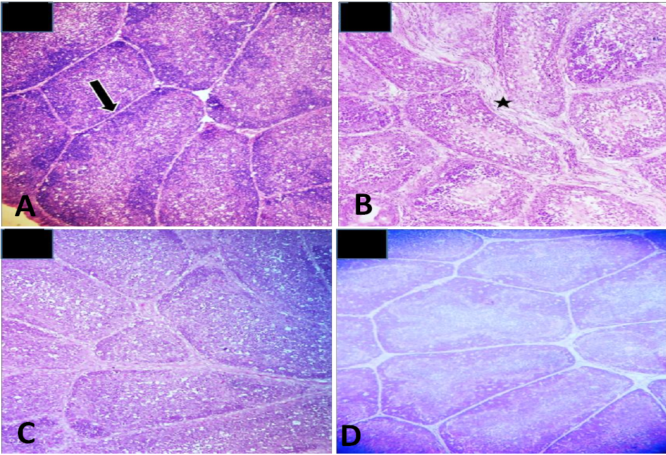
Fig. 3.
Histopathological examination of bursa of Fabricius of rats treated with aflatoxins and Bacillus licheniformis in different combinations. (A) Photomicrograph of group A (Basal diet) showing normal parenchyma of bursa of Fabricius. (B) Photomicrograph of group B (aflatoxins) showing increased interfollicular spaces. (C) Photomicrograph of group C (aflatoxins and Bacillus) showing almost normal parenchyma (D) Photomicrograph of group D (Bacillus) showing normal bursal parenchyma.