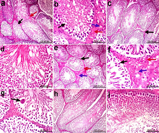
Representative photomicrographs of H & E stained sections of testes (scale bar, 100μm and 25 μm); a and b Fed on HFD; showing marked vacuolation of spermatogenic cells (black arrow) and Leydig cells (blue arrow) as well as interstitial oedema (red arrow). c and d Fed on HFD plus microcapsules of BL; showing normal seminiferous tubules with complete spermatogenic series and sperm production associated with slight intertubular oedema (black arrow). e, f and g Fed on HFD plus encapsulated SP and BL; e and f showing vacuolation of Leyding cells (blue arrow), marked vacuolation and necrosis of spermatogenic cells (black arrow) as well as interstitial oedema (red arrow); g) showing spermatid giant cell (black arrow). h and i Fed on basal diet (normal control) showing the normal histological architecture of seminiferous tubules with complete spermatogenic series and sperm production.